BISC 3754 Lecture Notes - Lecture 12: Secretion, Copii, Triskelion

28 Apr 2018
School
Department
Course
Professor

1
Intracellular Vesicular Traffic I
● Lumen of vesicle is the same as the extracellular region
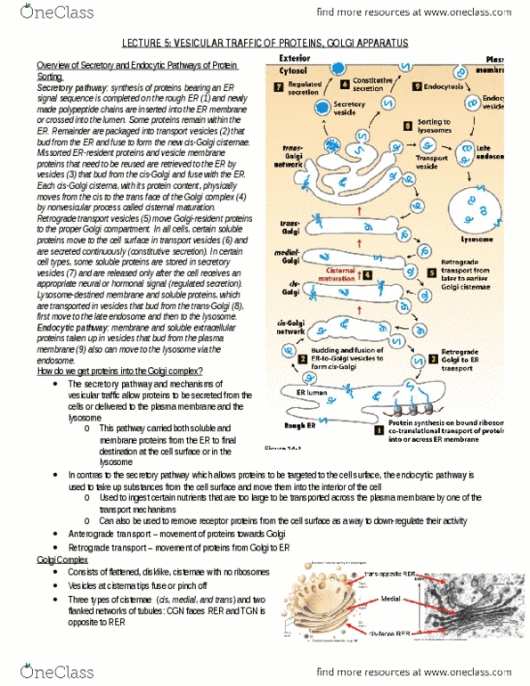
Exocytosis- transport vesicle fuses with plasma membrane, contents released into EC space
Endocytosis- plasma membrane internalizes, creates transport vesicle
Secretory pathway: rough ER → golgi → secretory vesicles → extracellular space
→ early endosome → extracellular space
→ late endosome → lysosome
Endocytic pathway- Plasma membrane to early endosome to late endosome to lysosome
● Molecular markers on cytosolic surface guide traffic
○ Allow transport vesicles to correctly fuse
○ Donor compartment membrane protein orientation is maintained
● Transport vesicles bud as coated= distinct protein coat on cytosolic side
○ Lost after it pinches off
○ Functions:
■ Inner coat layer selects appropriate mem molecules for transport
■ Outer coat layer shapes vesicle
● Clathrin coated- transport from golgi to plasma mem, between Golgi and endosome
● COPI/COPII- transport from ER to golgi
○ COPI- bud from golgi

2
○ COPII- bud form ER
● Vesicles need a coat protein
○ Has protein tag (recognition)
○ Cargo has protein binding sites/receptors
○ GTP binding protein- monomeric GTPase (small G protein)
Clathrin
● 3 large and 3 small polypeptide chains in each subunit= triskelion framework
● Adaptor proteins bind clathrin coat to membrane and trap proteins (between cage and mem.)
○ Adaptors are specific for cargo receptors
AP2 adaptor protein
● Each AP2 has 4 subunits and can bind to 4 PIP2
● Phosphoinositide P1(4,5)P2 in membrane (phospholipid)
○ AP2 binds to P1(4,5)P2→ changes conformation= binding sites for receptors exposed
● Cargo receptors also in membrane
○ Binding signals endocytosis
○ Cooperative process= more AP2 bind and coats are assembled once it starts
■ Due to curvature forming in membrane
● Phosphoinositides (PIP) → rapid phosphorylation/dephosphorylation
○ Distribution varies among organelles
○ PIP binding proteins regular vesicle formation and traffic
○ Reactions occur a 3, 4, or 5 carbon of sugar (inositol) head
Document Summary
Lumen of vesicle is the same as the extracellular region. Exocytosis- transport vesicle fuses with plasma membrane, contents released into ec space. Secretory pathway: rough er golgi secretory vesicles extracellular space. Endocytic pathway- plasma membrane to early endosome to late endosome to lysosome. Molecular markers on cytosolic surface guide traffic. Donor compartment membrane protein orientation is maintained. Transport vesicles bud as coated= distinct protein coat on cytosolic side. Inner coat layer selects appropriate mem molecules for transport. Clathrin coated- transport from golgi to plasma mem, between golgi and endosome. Gtp binding protein- monomeric gtpase (small g protein) 3 large and 3 small polypeptide chains in each subunit= triskelion framework. Adaptor proteins bind clathrin coat to membrane and trap proteins (between cage and mem. ) Each ap2 has 4 subunits and can bind to 4 pip2. Ap2 binds to p1(4,5)p2 changes conformation= binding sites for receptors exposed. Cooperative process= more ap2 bind and coats are assembled once it starts.