Physiology 1020 Lecture Notes - Lecture 8: Heart Valve, Atrioventricular Node, Purkinje Fibers

29 Sep 2017
School
Department
Course
Professor
Document Summary
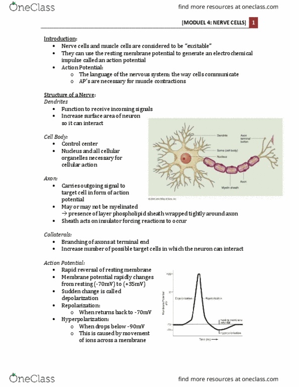
Form most of the walls of the atria and ventricles. Contain the same proteins actin and myosin arranged in bundles of myofibrils surrounded by a sarcoplasmic reticulum. Differ from skeletal muscle by having only 1 nucleus but far more mitochondria. 1/3 of their volume is taken up by mitochondria. Roughly 80% of the oxygen from the passing blood (twice the amount of other cells). Cells are much shorter, branched, and are joined together by special structures called intercalated discs. Gap junctions: allow for the movement of ions and ion currents between the myocardial cells. Because of the gap junctions, the myocardial cells of the heart can conduct action potentials from cell to cell without the need for nerves. Contract very weakly because they contain very few contractile elements (myofibrils). These cells are able to spontaneously generate action potentials without the help of nervous input like regular neurons. Can rapidly conduct the action potentials to atrial and ventricular muscle.