Anatomy and Cell Biology 3309 Lecture Notes - Lecture 6: Dense Irregular Connective Tissue, Superior Vena Cava, Aortic Valve

22 May 2018
School
Department
Professor

Histology 3309
The Heart
Learning Objectives
- Describe and identify the histological characteristics of the layers of the heart
- Describe the organization and histological characteristics of the pericardium
- Describe the location of the auto rhythmic cells of the heart and their role in propagating
in impulse through the heart
- Describe the histology of the heart valve
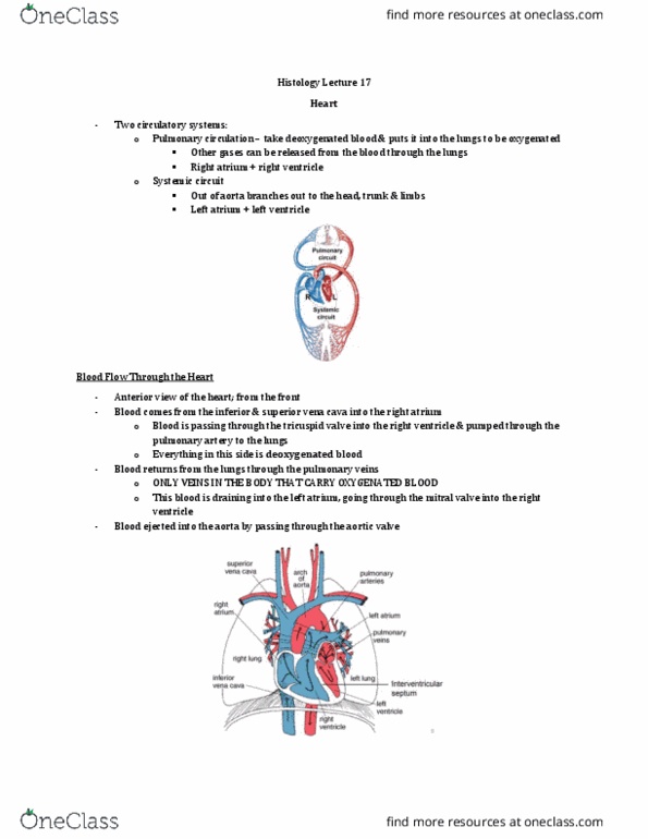
- There are 2 circulatory systems:
o Pulmonary circulation
▪ run off the right atrium and ventricle
▪ takes deoxygenated bloods and sends it to the lungs to become
oxygenated and to release other gases
o systemic circulation
▪ goes out through aorta to the rest of body
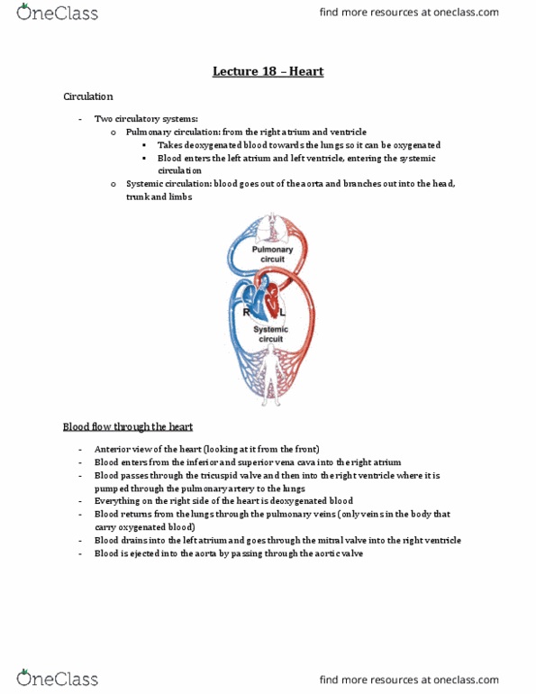
Blood Flow Through the Heart
- this is an anterior view of the heart (looking at it from the front)
- deoxygenated blood:
o blood is going to come from the inferior and superior vena cava into the right atrium
o that passes through the tricuspid valve into the right ventricle
o it is then pumped through the right artery into the lungs
- oxygenated blood
o blood returns from lungs through the pulmonary veins (only veins in the blood that
carry oxygenated blood)
o this drains into the left atrium and passes through mitral valve into the right ventricle
o this blood is ejected into the aorta by passing through the aortic valve
Fibrous Skeleton
- atria and the ventricles are separated from eachother by a connective tissue skeleton called
fibrous skeleton
- in the image, you see the plane cutting through at the level of the fibrous skeleton
- the atria have been removed – we are looking at the ventricles
- anterior is at the top, posterior is at the bottom
find more resources at oneclass.com
find more resources at oneclass.com

- the fibrous skeleton is dense irregular connective tissue
- is it nonconducting – acts as an insulator bw atria and ventricles
- it acts like a skeleton bc the cardiac muscle attaches to it – it takes origin from this fibrous
skeleton
- you can see the bundles of fibers running away from the fibrous skeleton
- the fibrous skeleton also acts as the point of attachment for the valves
- the valves that allow blood into the heart from the atria are the tricuspid valve (on right side)
and bicuspid valve (aka mitral valve) (on left side)
- blood comes out of the left ventricle through the aortic valve of the aorta and
- blood from the right ventricle goes up into the pulmonary artery through the pulmonary valve
-
Coverings of the Heart
- chamber of ventricle: where blood would be
- if we took off someones chest plate we would see mainly adipose tissue laid down on top of a
fibrous pericardium (dense irregular connective tissue)
o note: bovine heart valve replacement is made from the fibrous pericardium of a cow to
construct a valve to replace your own with when it stops functioning
- this fibrous pericardium has an inner lining towards the heart called the serous pericardium
o its made of mesothelial cells (a simple squamous epithelium)
o serous meaning it secretes fluid
- often we will combine the parietal layer of serous pericardium and fibrous pericardium and talk
about the parietal pericardium
- if we move towards the heart wall, we have another serous layer of the pericardium called the
visceral layer
- parietal and visceral refer to whether or not this pericardium is sitting on the organ (visceral) or
sitting away from the organ (parietal)
- in bw the parietal and visceral layers we have pericardial fluid
o allows heart to move without surfaces moving on one
another
o it’s a lubricant that prevents inflammation as the heart
pumps
o downside: if you have a wound to the heart that causes
blood to leak out, the parietal pericardium acts like a
barrier and the blood fills the pericardial cavity →the
heart can no longer relax and fill, its being squeezed by
the blood →need to get a catheter there and drain it
find more resources at oneclass.com
find more resources at oneclass.com

Pericardium
- this is with the chest plate removed
- heart is covered with parietal pericardium
- you see the aorta coming out and branches of the aorta (white lines)
- the darker lines are veins draining into the superior vena cava
- the surface of the heart has a shine on it and the parietal pericardium also has a shine on it
o the shine is the mesothelial layer
- how does this sac form? How do we get a sac that has mesothelium on the parietal and visceral
layer?
Serous Pericardium
- we have a balloon and you’re sticking your hand in it
- image that your forearm are the vessels that are entering and leaving the heart
- image that your fist is the heart
- if you stick your fist in an under inflated balloon, you are going to have a portion of that balloon
cover the fist and another portion reflected back to form a potential space
- the visceral pericardium is the part that is touching the fist (blue) and the parietal pericardium
is everything else (red)
- so the parietal and visceral pericardium are continuous with each other
- so the pericardium reflects back on itself – it goes from a visceral layer to a parietal layer
- and all of this is lined by a mesothelium which are producing the fluid you find in bw
find more resources at oneclass.com
find more resources at oneclass.com
Document Summary
Describe and identify the histological characteristics of the layers of the heart. Describe the organization and histological characteristics of the pericardium. Describe the location of the auto rhythmic cells of the heart and their role in propagating in impulse through the heart. Describe the histology of the heart valve. Blood flow through the heart this is an anterior view of the heart (looking at it from the front) Pericardium this is with the chest plate removed. Layers of the heart so there are 3 layers: epicardium. Non cardiac muscle layer that lines the chamber adjacent to blood supply one of the characteristics of blood is that its always in contact with endothelium (simple squamous epithelial) bc these cells are specialized to prevent coagulation from happening. Epicardium at top you can see the nuclei of the mesothelial cells (simple squamous epithelium) below hta tyou have fibroblasts and connective tissue.