Anatomy and Cell Biology 3309 Lecture Notes - Lecture 17: Superior Vena Cava, Tricuspid Valve, Pulmonary Valve
18 views11 pages

5 Apr 2018
School
Department
Professor
Document Summary
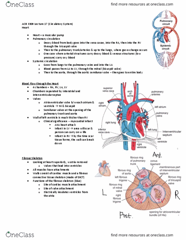
Anterior view of the heart; from the front. Blood returns from the lungs through the pulmonary veins: only veins in the body that carry oxygenated blood, this blood is draining into the left atrium, going through the mitral valve into the right ventricle. Blood ejected into the aorta by passing through the aortic valve. The atria & the ventricles are separated from each other by a connective tissue skeleton (fibrous skeleton) Plane cutting through at the level of the fibrous skeleton: atria have been removed, we are looking at the ventricle, anterior: top of diagram, posterior: bottom of diagram. Fibrous skeleton is: dense irregular connective tissue, non conducting; acts as an insulator between atria & ventricles. Chamber of the ventricle; where the blood is located. Adipose tissue laid down on top of a fibrous pericardium; outermost covering of the heart.
Get access
Grade+
$40 USD/m
Billed monthly

Homework Help
Study Guides
Textbook Solutions
Class Notes
Textbook Notes
Booster Class
10 Verified Answers
Class+
$30 USD/m
Billed monthly

Homework Help
Study Guides
Textbook Solutions
Class Notes
Textbook Notes
Booster Class
7 Verified Answers
Related Documents


Related Questions
Trace blood flow beginning with the left ventricle, going to the1st digit of the left foot, then back again.
use the word bank bellow
| Anterior tibial vein 2.Aortic semilunarvalve 3.Anteriortibialartery 4.Left ventricle 5.Bicuspidvalve 6.Tricuspidvalve 7.Pulmonaryarteries 8.Common iliac vein 9.Poplitealvein 10.Pulmonary veins 11.Left common iliacartery 12.Aortic arch 13.Inferior venacava valve 14.Alveolar capillaries 15. left atrium 16. Dorsalis pedicArtery 17. Ascending Aorta 18. Right ventricle 19. Femoral artery 20. Externaliliac vein 21. Thoracic aorta 22. Popliteal artery 23. Pulmonary trunk 24. Femoral vein 25. Abdominal aorta 26. Right atrium 27.Pulmonary semilunar 28. External iliac artery |
starts with 4, and end with 4 |


