1. Which of the following is the correct sequence of bloodflow?
a. heart --> atery --> arteriole --> capillary -->vein --> venule --> heart
b. heart --> vein --> venule --> capillary -->arteriole --> artery --> heart
c. heart --> artery --> arteriole --> capillary -->venule --> vein --> heart
d. heart --> artery --> venule --> capillary -->arteriole --> vein --> heart
e. heart--> artery --> capillary --> arteriole -->venule --> vein --> heart
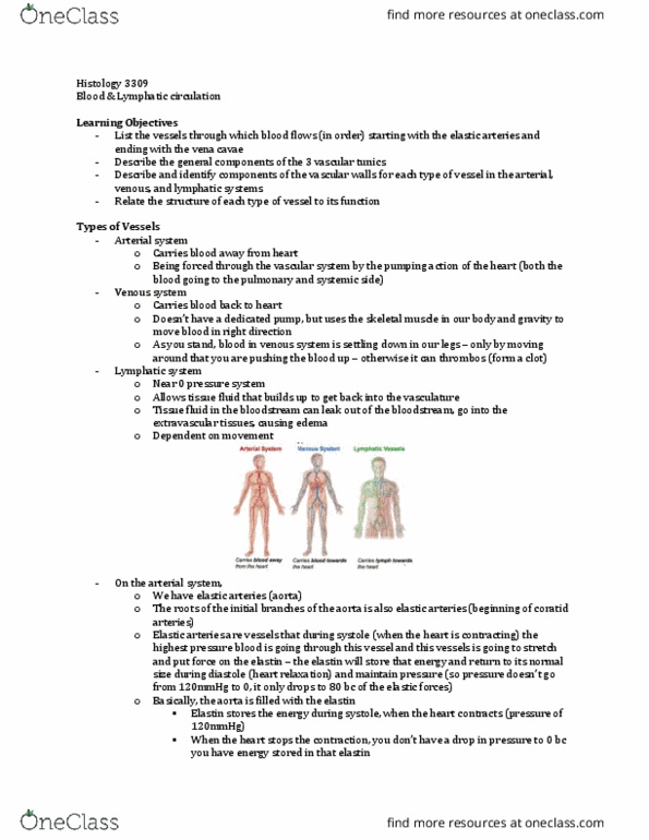
2. What are the functions of the elastic fibers that are foundin the walls of ateries? (there may be more than one answer)
Select one or more:
a. they allow the artery to stretch and then recoil back to itsoriginal shape as blood is forcegully pumped from the heart intothe arteries
b. they allow arteries to lengthen as the volume of blood in thearteries increase
c. they help maintain a steady blood pressure during thedifferent phases of heart contraction
d. they smooth out the walls of the arteries to preventturbulent blood flow
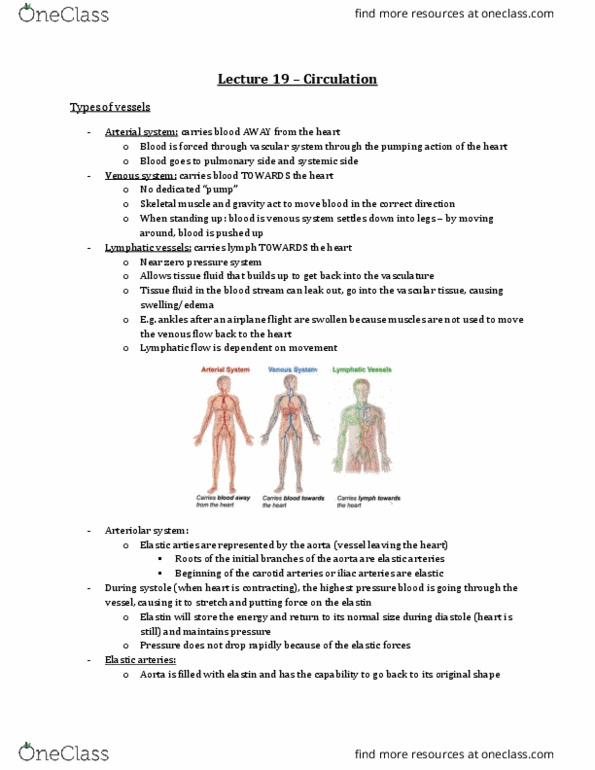
3. Which of the following are functions of the lymphatic system?(there may be more than one answer)
Select one or more:
a. help fight infections and cancer
b. allows excess interstitial fluid (fluid found between thecells in your tissues) to get dumped back into your blood
c. brings large molecules from fat digestion into thebloodstream so that they can be processed and stored
d. all of the above are functions of the lymphatic system
4. Which of the following blood vessels bring newly oxygenatedblood from the blood vessels in the lungs back to the heart so thatthe heart can pump it to the rest of the body?
Select one:
a. aorta
b. inferior vena cava
c. pulmonary vein
d. pulmonary artery
5. Which of the following mechanisms help blood fight gravity sothat, for instance, it can return from blood vessels in your feetback into your heart? (there may be more than one answer)
Select one or more:
a. breathing allows the pressure in your chest to decrease andallow blood to flow up towards your heart
b. as you walk, the skeletal muscles in your legs contract andsqueeze the veins; since veins have valves, blood flows up anddoesn't backflow back down
c. the force of blood flowing out of the heart and into thearteries pushes all of the blood along, causing blood to return tothe heart
d. precapillary sphincters open to allow blood to flow backtowards the heart
6. The tricuspid and bicuspid/mitral valves prevent backflow ofblood from the atria into the ventricles.
Select one:
True
False
7. The following sequence represents the correct order of bloodflow through the heart:
inferior and superior vena cava --> right atrium --> rightventricle --> pulmonary artery --> lungs --> pulmonaryvein --> left atrium --> left ventricle --> aorta
Select one:
True
False
8. Capillaries are as wide as one red blood cell.
Select one:
True
False
9. The purpose of the coronary arteries is to bring oxygenatedblood to the walls of the heart so that the muscles in the heartwalls can contract.
Select one:
True
False
10. Vasodilation refers to the narrowing of blood vessels.
Select one:
True
False