BIO130H1 Lecture Notes - Lecture 26: Spindle Apparatus, Cell Division, Kinetochore

4 Apr 2019
School
Department
Course
Professor

BIO130H1 verified notes
26/26View all
Document Summary
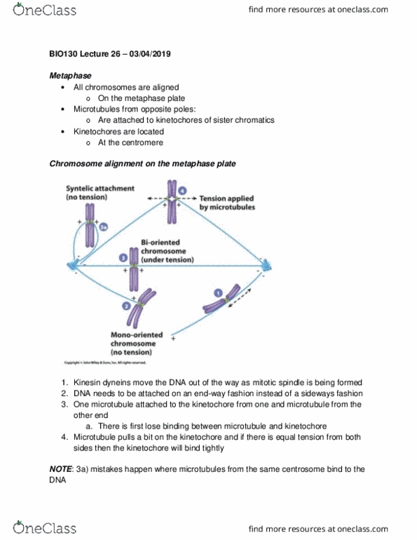
Metaphase: all chromosomes are aligned, on the metaphase plate, microtubules from opposite poles, are attached to kinetochores of sister chromatics, kinetochores are located, at the centromere. Note: 3a) mistakes happen where microtubules from the same centrosome bind to the. They pull in the same direction causing the kinetochore to not bind tightly to the microtubule. This leads to the disassembly of the microtubule (depolymerization) Chromosomes attachment to microtubules: exposed plus end of the microtubule allows for polymerization / depolymerization, the kinetochore binds near the plus end of the microtubule but not directly on the plus end! Tubulin flux through microtubules: centrosome nucleation of microtubules. If red radioactive tubulin dimers are added, the polar microtubules and kinetochore microtubules become red: this is because there is microtubule treadmill. In the centrosome there are special depolymerases that remove heterodimers one at the time: microtubule treadmilling, there depolymerases are normally not on the centrosomes, they are found only during metaphase.