MEDRADSC 3C03 Lecture Notes - Lecture 9: Renal Pelvis, Renal Cortex, Renal Pyramids

8 Jun 2020
School
Department
Course
Professor
Document Summary
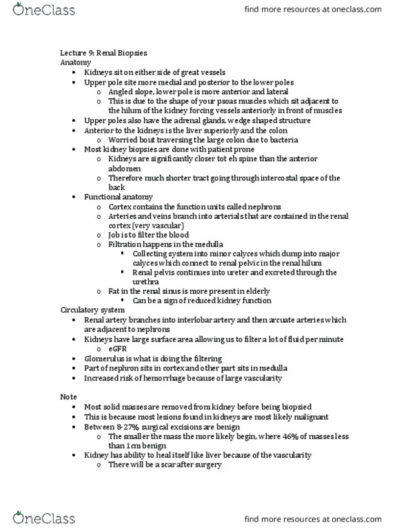
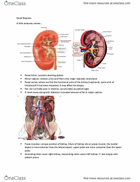
Anatomy review - what to keep in mind when planning a biopsy track. Upper pole sits more medial & posterior than lower - sit on an oblique plane. Bc of the psoas muscles - sit adjacent to hilum. Hilum is turned slightly anterior bc of the muscles. Great vessels sit slightly anterior to the kidneys. Location of liver (for r kidney) & ascending colon (r kidney), transverse colon, & descending colon (l kidney) Most kidney biopsies are done with patient prone for this reason - access kidneys through intercostal spaces. Shows hilum sitting more anterior - towards the great vessels (cortex sits more posteriorly) Arteries & veins branch out into tiny arterioles contained within renal cortex. Renal pyramid (medulla) - area where filtration happens. Waste & excess water from blood - excreted into collecting system - into renal calyces - into major calyces - collect into renal pelvis in the hilum - connects into ureter.