ZOO 3733C Study Guide - Quiz Guide: Right Triangular Ligament, Common Hepatic Duct, Ductus Venosus

Document Summary
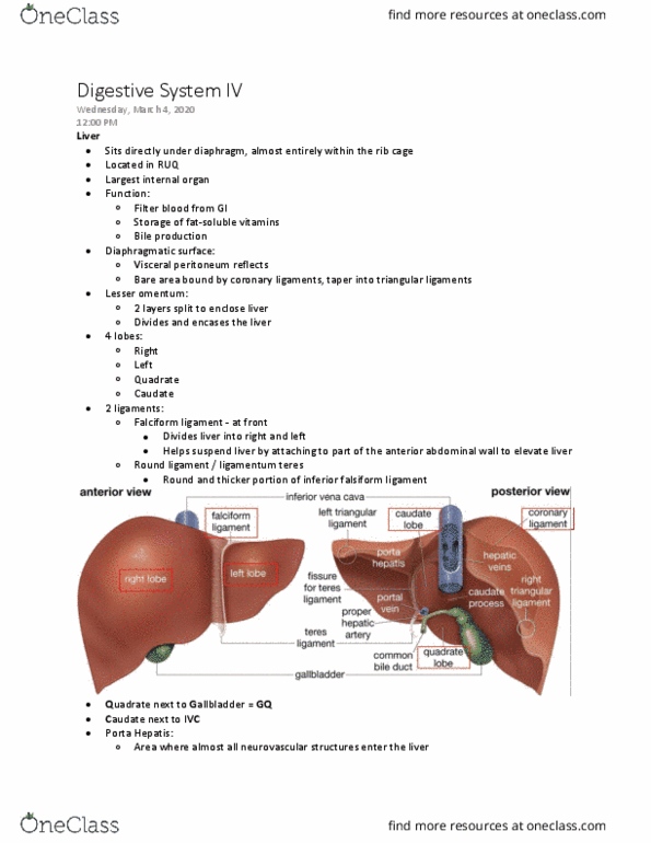
Peritoneum clings to abdominal wall and organs: parietal: portion that lines abdominal and pelvic cavities, visceral: portion that covers external surfaces of most abdominal organs. Ligamentum venosum in fetal life is ductus venosus. Round (aka teres) ligament in fetal life is l umbilical v. Right coronary ligament-white diving line: becomes right triangular ligament. Left coronary ligament becomes left triangular ligament. Caudate lobe- next to ivc (posterior side) Quadrate lobe- next to gallbladder, square-shaped (posterior side) Hepatic- liver: l&r hepatic ducts -> common hepatic duct -> gallbladder -> cystic duct -> cbd. Portal triad aka glistens triad: purple- portal vein, red- hepatic artery (proper, green- cbd. Bare area interacts with diaphragm- only area not covered by peritoneum. One of its main jobs is the recreation of digestive enzymes. Split o of main pancreatic duct ( sh bone-looking structure): accessory pancreatic duct.