BIOC 4580 Study Guide - Midterm Guide: Halophile, Palmitoylation, Ras Superfamily

12 Feb 2017
School
Department
Course
Professor
Document Summary
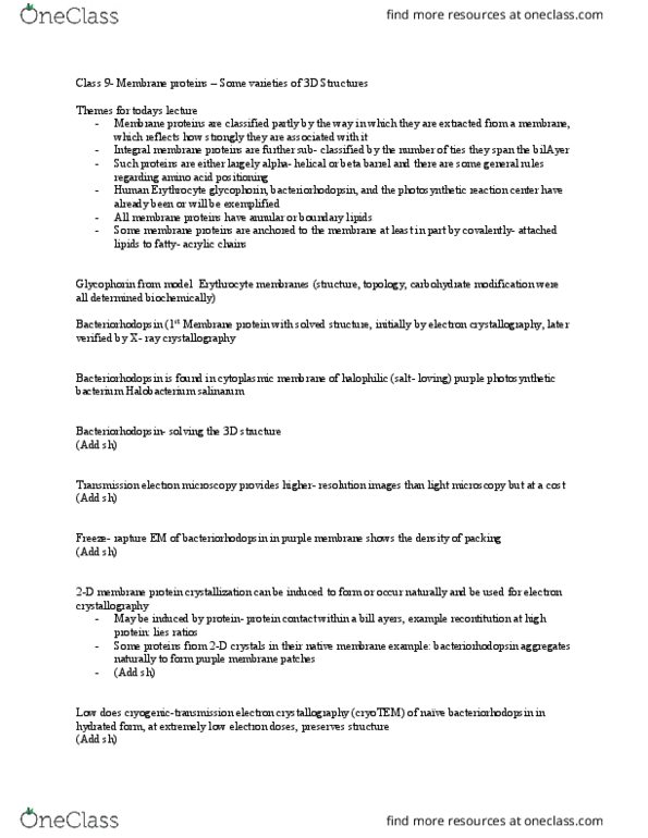
Lecture 7: membrane proteins- some varieties of 3d structures. Bacteriorhodopsin found in the cytoplasmic membrane of halophilic purple photosynthetic bacterium. All charged residues are on one face of each helix, facing inside helix bundle. Easy to study (cid:271)e(cid:272)ause it is so highly pa(cid:272)ked that it (cid:862)(cid:272)rystallizes itself(cid:863) Does not need to be purified and reconstituted. 2d membrane protein crystallization you can combine 2d images to create a 3d structure. Monomers have 7 membrane-spanning alpha-helices 23 amino acids each. Forms a trimer with hydrophobic residues pointing out and hydrophilic ones pointing in to form a channel. In channel, retinal (pigment) covalently bound to lys216 residue. Retinal switched from trans to cis form upon photon absorption. Upon isomerization, a proton (h+) is passed to an asp residue which passes it on to the extracellular space. Another asp accepts h+ from cytoplasm, passes it to retinal, cycle begins again. Proton gradient formed by bacteriorhodopsin used to drive production of atp.