IMED3001 Study Guide - Final Guide: Coronary Sinus, Pulmonary Artery, Bulbus Cordis

14 Dec 2020
School
Department
Course
Professor
Document Summary
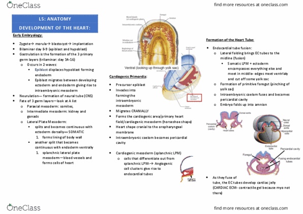
The single heart tube is symetrical with paired inflow and outflow tubes. It is endocardium, myocardium with a pericardial coelom or sac around it. Above: formation of the s-shaped heart from fused cardiac tubes in the human embryo at about 21 to 23 days. First part of body to show signs of asymmetry. Lower part tucks up" under the cranial part. The distal or cranial part of the loop then swings to the right (s- shaped) The caudal part shifts dorsally to come to lie behind the outflow tract. The bulbus cordis becomes the conus arteriosus and truncus arteriosus aortic sac and arch system. The middle loop of the s becomes the r ventricle, the more caudal part. Becomes the l ventricle, and finally the atrial part becomes. Ventral views of human embryonic hearts that illustrate bending of the cardiac tube and the establishment of its regional divisions. Smooth part of r ventricle (conus arteriosus)