ANHB2212 Lecture Notes - Lecture 12: Endocardial Cushions, Interatrial Septum, Ectoderm

28 May 2018
School
Course
Professor

L12 Heart Development
Outcomes
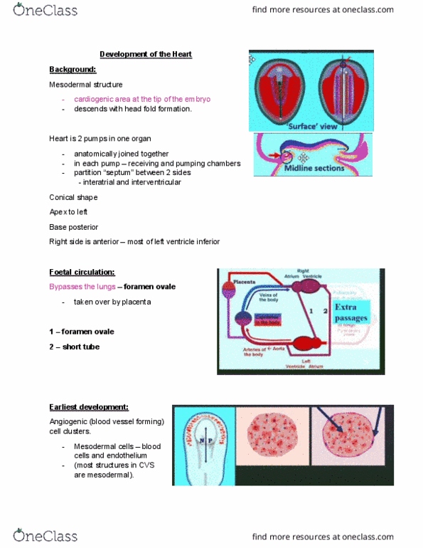
Foetal circulation
Lungs are not functional prior to birth
Blood from RV has no where to go – as lung is not open
Oxygen comes from placenta
Blood must go to the Left side
RV has to develop to pump some blood because if structures
have no blood, they die out
1. Blood flows from RA to LA (hole)
2. Pulmonary Artery is close to Aorta
p5
Cardiogenic area on trilaminar embryo
• There is a section without mesoderm, where
Ectoderm and Endoderm are in contact
o This is the Cardiogenic area
• There is a section at head end
o This is the Oropharyngeal membrane
p7
Cranial end (i.e arterial end)
Caudal end (i.e venous end
Starting from caudal end
1. Sinus Venosus
2. Atrium
3. Ventricle
4. Bulbus
5. Conus
6. Truncus arteriosus (gives rise to Aortic Arches (3))
Ends at Cranial end
Tube bends
• Atrium (1) + Sinus Venosus (posterior)
• Ventricle (1, no R/L) + Bulbus (anterior)
• V + B form Bulboventricular Loop
o Ventricles to the Left
o Bulbus to the Right
Interior
• Single atrium joined at the posterior
• Common Atrioventricular canal (1→ 1)
• Outflow from ventricles/bulbus (1)
Forming 4 chambers from 2 chambers by septa
Recall: Interventricular, Interatria
1. Interatrial septum
• Forms atrioventricular division
o With endocardial cushions (AV cushions)
o 1 atrium → 2 atria
2. Interventricular septum
• splitting conus and truncus
o with spiral aortic septum (L)
o with spiral pulmonary septum (R)
find more resources at oneclass.com
find more resources at oneclass.com
Document Summary
Blood from rv has no where to go as lung is not open. Rv has to develop to pump some blood because if structures have no blood, they die out. Cardiogenic area on trilaminar embryo: there is a section without mesoderm, where. Ectoderm and endoderm are in contact: this is the cardiogenic area, there is a section at head end, this is the oropharyngeal membrane p7. Starting from caudal end: sinus venosus, atrium, ventricle, bulbus, conus, truncus arteriosus (gives rise to aortic arches (3)) Tube bends: atrium (1) + sinus venosus (posterior, ventricle (1, no r/l) + bulbus (anterior, v + b form bulboventricular loop, ventricles to the left, bulbus to the right. Interior: single atrium joined at the posterior, common atrioventricular canal (1 1, outflow from ventricles/bulbus (1) Forming 4 chambers from 2 chambers by septa.