BIO 365S Chapter Notes - Chapter 4: Atrioventricular Node, Flowchart, Ventricular Fibrillation


Discussion Exercise—ECG
1. Draw a flowchart illustrating the normal impulse conduction through the heart. How is the
depolarization conveyed from cell to cell?
2. An electrocardiogram is a sum of all the electrical activity of the heart, recorded from the
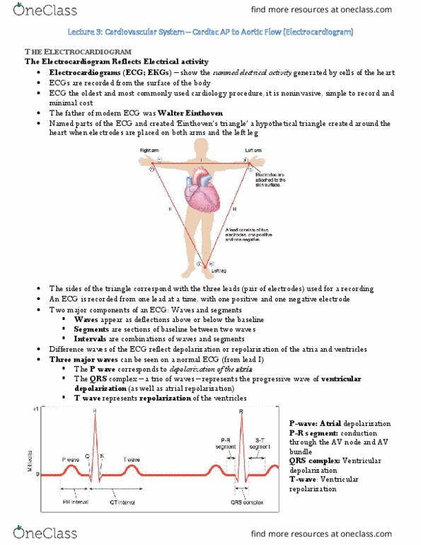
surface of the body. For the normal ECG below:
a. Label the waves and segments.
b. Calculate the heart rate.
a. 4 heart beats per 3 sec
b. to find heartbeats in 60 sec, set up proportion
c. (4 beats/3sec) = (x beats/60 sec)
d. **Where the line is = 1 beat. Common mistake, people would think this is 2.
c. Which wave, segment or interval would you analyze to determine:
AV node delay same about of time b/w beats; Analyze p waves. PR segment.
Atrial rhythm
Conduction through ventricular muscle QRS
Refractory period of contractile AP time b/w QRS and T.
SA node rhythm = P waves.
d. In which part of the ECG does the ventricular Na+ influx occur?
3 a. .How would the trace in #2 change under sympathetic influence on pacemaker cells?
b. Describe the cellular mechanisms that lead to those changes.
3 sec
find more resources at oneclass.com
find more resources at oneclass.com
Document Summary
Discussion exercise ecg: draw a flowchart illustrating the normal impulse conduction through the heart. How is the depolarization conveyed from cell to cell: an electrocardiogram is a sum of all the electrical activity of the heart, recorded from the surface of the body. Common mistake, people would think this is 2: which wave, segment or interval would you analyze to determine: Av node delay same about of time b/w beats; analyze p waves. Sa node rhythm = p waves: in which part of the ecg does the ventricular na+ influx occur? time b/w qrs and t. how would the trace in #2 change under sympathetic influence on pacemaker cells: describe the cellular mechanisms that lead to those changes. 5a. the following strips illustrate a fairly common pathology. What is the difference between the recording in a versus b? a. Identify the above two traces as v-fib (ventricular fibrillation) or a-fib (atrial fibrillation).