Anatomy and Cell Biology 3319 Chapter Notes - Chapter 1-30: Median Aperture, Ependyma, Lateral Ventricles

13 Apr 2015
School
Department
Professor
Document Summary
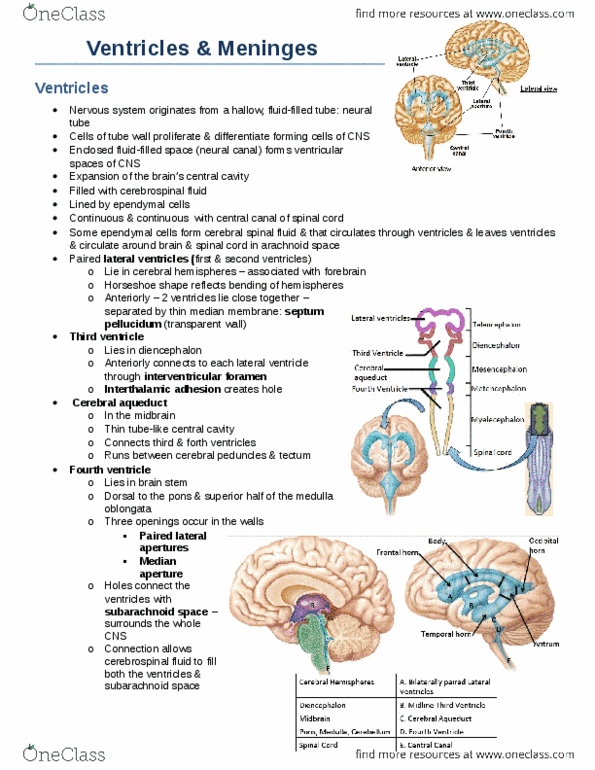
Ventricles ventricles are expansions of the brains central cavity which contain csf and are lined by ependymal cells. The lateral ventricles (1/2) lie within the cerebral hemispheres (horseshoe shape: they lie close to each other and are only separated by a thin membrane called the septum pellucidum. The third ventricle lies in the diencephalon and connects to the lateral ventricle through the interventricular foramen. The cerebral aquedeuct is found in the midbrain and is a tube central cavity which connects the 3 and 4 ventricle. The dura mater is a two layered sheet of fibrous connective tissue: the more superficial layer is the periosteal layer which attaches to the internal surface of the skull, the deeper meningeal layer forms the external covering of the brain, the two layers are fused except when they enclose the blood filled dural sinuses. of the neck.