Anatomy and Cell Biology 3319 Study Guide - Midterm Guide: Vagus Nerve, Infraorbital Foramen, Zygomatic Arch

22 Mar 2018
School
Department
Professor
Document Summary
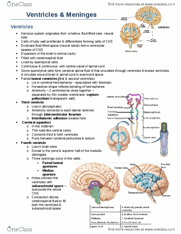
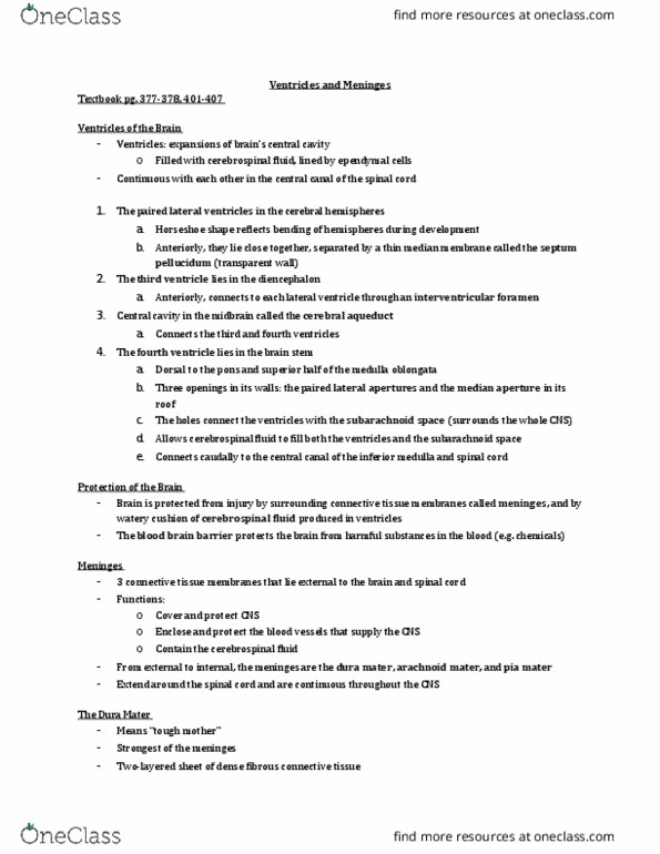
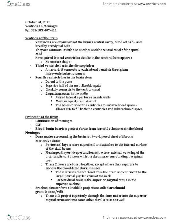
A(cid:374)ato(cid:373)(cid:455) (cid:1007)(cid:1007)(cid:1005)9: test (cid:1006) tud(cid:455) heet (lectures 12 21) Formation of cerebral spinal fluid: formed by specialized cells in choroid plexus. Expands to various regions (i. e. forebrain, midbrain, etc. ) Eventually go on to form different parts of the brain. The spaces that were originally in the interior part of the neural tube remain as spaces (i. e. ventricles) Lateral ventricle, third ventricle, fourth ventricle, then everything gets narrowed down and continues on to the spinal cord. Note: lateral ventricles are ventricles 1 and 2; they are the largest ventricles. Slides (left) show where the ventricles will be. Third and fourth ventricle connected by cerebral aqueduct. Fourth ventricle ends up becoming the central canal. All ventricles are continuous (a) just barely grazes the lateral ventricles; only see a little bit of the ventricle; if we went a little bit deeper, we could see a bit more of the lateral ventricles (b) Choroid plexus are cells that line the ventricles.