MEDI7302 Study Guide - Final Guide: Esophagus, Posterior Auricular Artery, Anterior Ramus Of Spinal Nerve


Neck Region
Learning
objectives
Understand basic topographical anatomy of the neck, including CT anatomy
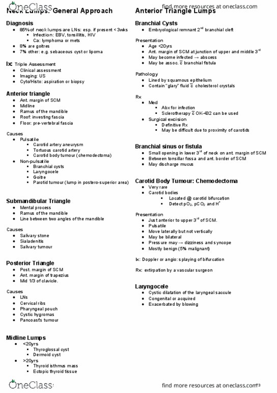
Describe the embryologic descent and fate of the thyroglossal tract
Explain the origin and presentation of branchial cysts
Recognize the fundamental differences between benign and malignant lumps within
the salivary glands
Describe the presentation and management of submandibular salivary calculi
Describe the pathophysiology of a pharyngeal pouch
Outline how to clinically recognize a carotid body tumour
Outline the diagnostic approach to a pathologic lymph node in the neck
Anatomy Neck is a conduit between head and body
Overall
Areas Anterior
triangle
Borders - midline neck (medial),
sternocleidomastoid (lateral), mandible (superior), investing
fascia (roof), visceral fascia (floor)
Contents
Suprahyoid muscles - mylohyoid,
geniohyoid, digastric, stylohyoid
Infrahyoid - sternohyoid,
sternothyroid, thyrohyoid, omohyoid
Common carotid artery bifurcation -
EC and IC branches
Internal jugular vein
CN VII (facial), CN IX
(glossopharyngeal), CN X (vagus), CN XI (accessory), CN XII
(hypoglossal)
4 smaller triangles inside anterior triangle
Carotid triangle
find more resources at oneclass.com
find more resources at oneclass.com

Submental triangle
Submandibular triangle
Muscular triangle
Posterior
triangle
Boundaries - sternocleidomastoid (anterior),
trapezius muscle (posterior), middle 1/3 clavicle (inferior),
investing fascia (roof), prevrtebral fascia (floor)
find more resources at oneclass.com
find more resources at oneclass.com

Contents
Omohyoid, vertebral muscles
(splenius capitis, levator scapulae, scalenes)
External jugular vein, subclavian
vein, transverse cervical and suprascapular vein
Subclavian artery, transverse cervical
artery, suprascapular artery
Accessory nerve (CN XI), cervical
plexus (phrenic nerve C3-5), brachial plexus
Fascia Superficial cervical (between dermis -> deep cervical
fascia)
Skin neurovascular supple
Superficial veins (eg external jugular)
Superficial lymph nodes
Fat
Platysma muscle
Deep cervical (underneath superficial fascia; several
layers)
Investing layer (most superficial)
oSurrounds all structures in neck, including
trapezius and sternocleidomastoid
Pre-tracheal (anterior neck)
oVisceral contents - oesophagus, thyroid,
trachea
oMuscular contents - infrahyoid muscles
Pre-vertebral (vertebral area)
oSurround vertebral column + associated
muscles (scalene, pre-vertebral, deep muscles of back)
oSurrounds brachial plexus + subclavian
artery to form axillary sheath
Carotid sheath
oCommon carotid + bifurcation into EC and
IC arteries
oInternal jugular vein
oVagus nerve
oCervical lymph nodes
find more resources at oneclass.com
find more resources at oneclass.com
Document Summary
Understand basic topographical anatomy of the neck, including ct anatomy. Describe the embryologic descent and fate of the thyroglossal tract. Explain the origin and presentation of branchial cysts. Recognize the fundamental differences between benign and malignant lumps within the salivary glands. Describe the presentation and management of submandibular salivary calculi. Outline how to clinically recognize a carotid body tumour. Outline the diagnostic approach to a pathologic lymph node in the neck. Neck is a conduit between head and body. Borders - midline neck (medial), sternocleidomastoid (lateral), mandible (superior), investing fascia (roof), visceral fascia (floor) Cn vii (facial), cn ix (glossopharyngeal), cn x (vagus), cn xi (accessory), cn xii (hypoglossal) Boundaries - sternocleidomastoid (anterior), trapezius muscle (posterior), middle 1/3 clavicle (inferior), investing fascia (roof), prevrtebral fascia (floor) Omohyoid, vertebral muscles (splenius capitis, levator scapulae, scalenes) External jugular vein, subclavian vein, transverse cervical and suprascapular vein. Accessory nerve (cn xi), cervical plexus (phrenic nerve c3-5), brachial plexus.