LIFESCI 23L Lecture Notes - Lecture 9: Pap Test, In Situ Hybridization, Teratoma


Lecture I: Histology
Overview of concepts and skills
● History and histology and staining techniques
● Dissecting vs. compound microscopes
● Performing a histology lab exercise w/ aid from computer tutorial
● Relate structure to function of cells and tissue
● SKILLS: proper use of dissecting and compound microscopes and proper handling of slides
● CONCEPTS: making connection between morphology and function
Background Information
● Gross anatomy → macroscale anatomy (studied last week)
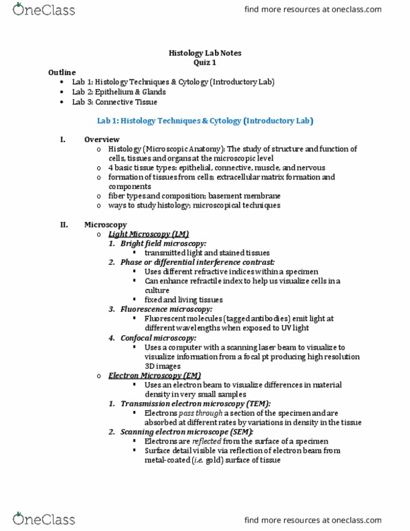
● Histology = study of microscopic anatomy of cells and tissues
○ Important method of analyzing tissues
○ Came about after the invention of the microscope
○ Various dyes and staining techniques used to visualize tissues
■ Microscopic structure of cells, tissues, and organs are all relate dto their functions
■ (Masson’s) Trichrome stain
● Cytoplasm = pink, keratine/muscle fibers = red, collagen/bone = blue; nuclei =
black
■ Haematoxylin (H&E → used in lab)
● Composed of two separate dyes; attracted to acids like nucleic acids (acidophile)
● Stains nuclei dark purple
■ Eosin
● Basophile, stains alkaline parts of the cell such as cytoplasm
● Counterstain → stains background and adds contrast
● Histopathology = histology used to diagnose disease, but also as a preventative method and in
research
○ E.g. collecting biopsy sample from suspicious site and viewed under microscope (e.g. potential
tumor cells)
○ Pap smear → preventitive method against cervical cancer
○ Colonoscopy → preventative method against colon cancer
○ Teratoma assay → suspected stem cell is injected into a mouse to see if a teratoma forms
■ Teratomer → tumor w/ many different kind of tissue; used to see if stem cell is pluripotent
(able to differentiate into tissues from all 3 germ layers)
● Histology used to analyze teratoma
● Each tissue has unique appearance under microscope → can be identified w/
histology
○ Immunohistochemistry → antibodies used to mark specific proteins or other substances in a
tissue slice
■ Antibodies are linked to enzymes, then a substrate is added to the slide
■ Enzyme cleaves substrate into colorful stain that can be seen under microscope
○ In situ hybridization → labelled DNA or RNA probe is used to detect specific mRNA’s in a cell
○ Bainbow → mapping connections in the brain
find more resources at oneclass.com
find more resources at oneclass.com