BIO 115 Lecture Notes - Lecture 15: Chest Radiograph, Trachea, Descending Aorta

Document Summary
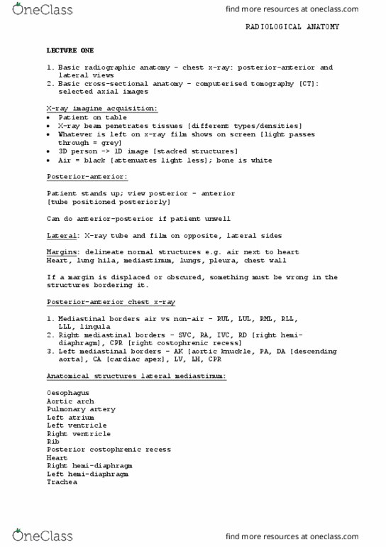
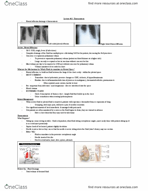
Radiologic lung anatomy: describe a systematic approach to reading a chest radiograph based on an understanding of the normal anatomy, focusing on the trachea/mainstem bronchi, hila, pleura, and lung parenchyma. Mks1a: costophrenic angles - the sharp, lateral, inferior extensions of the lungs, blunting is caused by pleural effusions, explain the concept of a pleural recess and describe at which thoracic level the pleura descends posteriorly, laterally, and anteriorly. Mks1a: pleura extends to the level of t12 posteriorly, t10 laterally, t8 anteriorly, explain the concept of the silhouette sign and why it is important for localizing structures in radiology. Know the location of the different lobes on pa and lateral chest radiographs. Some abnormalities on chest x-ray based on alteration of the normal anatomy. (right main bronchus intubation, lobar pneumonia, pleural effusion, pneumothorax, edema, hilar enlargement). Mks1b lobar pneumonia - opaque densities on film. look for silhouette as well. Right main bronchus intubation - down the wrong bronchus at the carina.