BIO 361 Lecture Notes - Lecture 17: Antibody, Glycosylation, Reduction Potential

29 Jun 2018
School
Department
Course
Professor

BIO361.30 & .60
Discussion Clarification Week 4, Post 5, Lectures 17-21
Lecture 17: Effects of carboxypeptidases on hemoglobin
1. Carboxypeptidase A can be used to remove the last two residues from the beta chains of
hemoglobin A. This should result in:
A. loss of two of the four oxygen binding sites.
B. a reduced magnitude of the Bohr effect without any significant reduction in the total oxygen
binding capacity (the numbers of oxygens bound at saturating O2 pressures).
C. a protein that has increased stability of the low affinity conformation.
D increased oxygen affinity without any change in the Bohr effect.
E. a protein that should have increased affinity for 2,3-DPG or IHP.
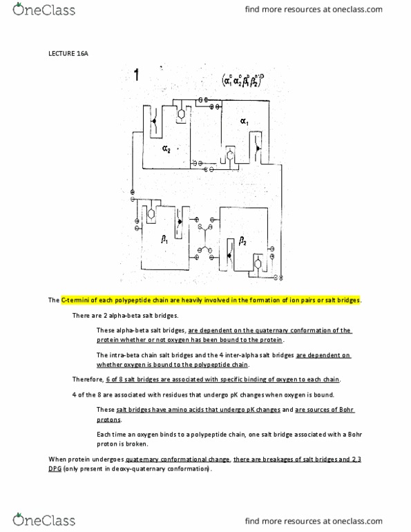
The effects of O2 binding onto the heme groups of individual deoxygenated hemoglobin (Hb)
subunits can be described as the progressive breaking of eight so-called “oxygenation-linked”
salt bridges involving the C-termini of the subunits. These salt bridges each help stabilize the
deoxygenated form of Hb. Of these eight salt bridges, only four involve amino acids (either N-
terminal amino groups or imidazole side chains) that hold onto a “Bohr proton” specifically
when participating in a salt bridge when in the deoxygenated form. The C-terminal histidines as
well as the adjacent “penultimate” tyrosines of the beta chains can be removed with
Carboxypeptidase A (the expopeptidase can remove all residues except for lysine and arginine).
The phenolic side chain of the penultimate tyrosine residue fits into the FG corner of each beta
chain when the subunit is deoxygenated. As long as the tyrosine residue remains in the FG
corner, the histidine’s imidazole group is within close proximity to a deprotonated aspartic acid
side chain, which has the effect of increasing histidine’s pK, allowing it to hold onto a Bohr
proton. However, binding of O2 to a heme group of a beta subunit results in a conformational
change that expels the tyrosine residue from the FG corner, dragging its histidine neighbor away
from the aspartic acid residue. This results in a decrease in histidine’s pK, and subsequent release
of its Bohr proton; this is in essence the molecular basis of the Bohr effect, at least for the beta
chains. Loss of these residues would therefore result in an observed decrease of the Bohr effect
due to destruction of the corresponding salt bridges and an apparent increase in the affinity of the
hemoglobin tetramer for oxygen, but because the structures of the oxygen-binding sites (the
heme groups) are preserved, the actual maximum binding capacity of Hb would not be affected.
Choice B is therefore the answer. It is important to note that binding of 2,3-DPG to the cavity
between the two beta-chains of Hb can only occur in the deoxygenated form; the cavity is too
small when Hb is oxygenated.
Lecture 18: MWC v. KNF models
2. You are trying to decide whether the ligand-binding characteristics of a tetrameric protein are
best described by the Monod-Wyman Changeux model or the Koshland- Nemethy-Filmer model
of cooperativity. Which observation fits the conclusion?
find more resources at oneclass.com
find more resources at oneclass.com

A. If the value of the Hill constant for ligand binding is 0.5, the Monod-Wyman Changeux
model is supported.
B. If you have two different antibodies that can detect the two different conformations of the
protein and both antibodies recognize the protein regardless of whether it has ligands bound, the
Koshland-Nemethy-Filmer model is ruled out.
C. You have a sensitive machine that can quantitate what fraction of the protein has undergone a
conformational change and another machine that can quantitate what fraction of the protein has
ligand bound. The two measurements don’t give the same value when you partially saturate the
protein with ligand. These observations are best explained by the Koshland-Nemethy Filmer
model.
D. You have discovered a low molecular weight molecule that affects the affinity of the protein
for ligand. Structural analysis shows that this small molecule stabilizes the protein in a single
conformation that has high affinity for ligand, but the small molecule does not bind anywhere
near the ligand. This result is most easily explained by the Koshland- Nemethy-Filmer model.
E. The protein releases protons into the medium when ligand is bound. This proton release
appears to be strictly linearly related to the fractional saturation with ligand. This observation is
best accommodated by the Monod-Wyman-Changeux model.
The Monod-Wyman-Changeux (MWC) and Koshland-Nemethy-Filmer (KNF) models are two
distinct models for cooperativity in proteins with more than one subunit. The MHC model, often
referred to as the “concerted” model, describes only two conformations for the entire mulitmeric
protein, a relaxed (R) conformation with relatively high ligand affinity, and a constrained (T)
conformation with lower ligand affinity. The two conformations are always in equilibrium with
each other (i.e., some of the protein molecules are always in each form), and the position of the
equilibrium shifts depending on how many subunits have ligand bound. Due to the model’s
inherent simplicity, the ligand-binding curve can be described using only two parameters, the
ratio of each conformation’s affinity for ligand (“c”, equivalent to KR, the dissociation
equilibrium constant for the relaxed form, divided by KT, the dissociation equilibrium constant
for the constrained form), and the value of the conformational equilibrium constant for the two
conformations when no ligand is bound (“L”). The KNF model, colloquially referred to as the
“sequential” model, instead describes two conformations for each subunit of the protein,
depending on whether or not each subunit has ligand bound. These ligation-linked
conformational changes within each subunit are obligate (there is no equilibrium between
conformations in the absence of substrate binding) and are always confined to the subunit that
has bound ligand. The conformational changes that occur in each subunit when ligand binds
result in changes in inter-subunit contacts that lead to free energy changes that can promote or
inhibit binding of ligands to the neighboring subunits. Therefore, depending on the alterations to
the inter-subunit contacts, the KNF model can give rise to either a sigmoidal ligand-binding
curve that displays positive cooperativity or, alternatively, a flattened hyperbolic curve that
displays negative cooperativity. Each model has its own limitations. The MWC model, because it
only allows for two overall conformations, can only explain positive cooperativity or non-
cooperativity, as negative cooperativity would require a third conformation with affinity lower
than the constrained (T) form. Therefore, the Hill constant (nH) can never be less than 1
according to the MWC model. Only the KNF model can easily explain negative cooperativity.
However, the MWC model can easily explain allosteric effectors like 2,3-DPG that bind
exclusively or selectively to one of the two quaternary conformations of hemoglobin as causing a
find more resources at oneclass.com
find more resources at oneclass.com
Document Summary
Discussion clarification week 4, post 5, lectures 17-21. Lecture 17: effects of carboxypeptidases on hemoglobin: carboxypeptidase a can be used to remove the last two residues from the beta chains of hemoglobin a. D increased oxygen affinity without any change in the bohr effect: a protein that should have increased affinity for 2,3-dpg or ihp. The effects of o2 binding onto the heme groups of individual deoxygenated hemoglobin (hb) subunits can be described as the progressive breaking of eight so-called oxygenation-linked salt bridges involving the c-termini of the subunits. These salt bridges each help stabilize the deoxygenated form of hb. Of these eight salt bridges, only four involve amino acids (either n- terminal amino groups or imidazole side chains) that hold onto a bohr proton specifically when participating in a salt bridge when in the deoxygenated form. The c-terminal histidines as well as the adjacent penultimate tyrosines of the beta chains can be removed with.