NUR 306 Lecture Notes - Lecture 4: Hyperthyroidism, Aortic Stenosis, Pericardial Effusion

Document Summary
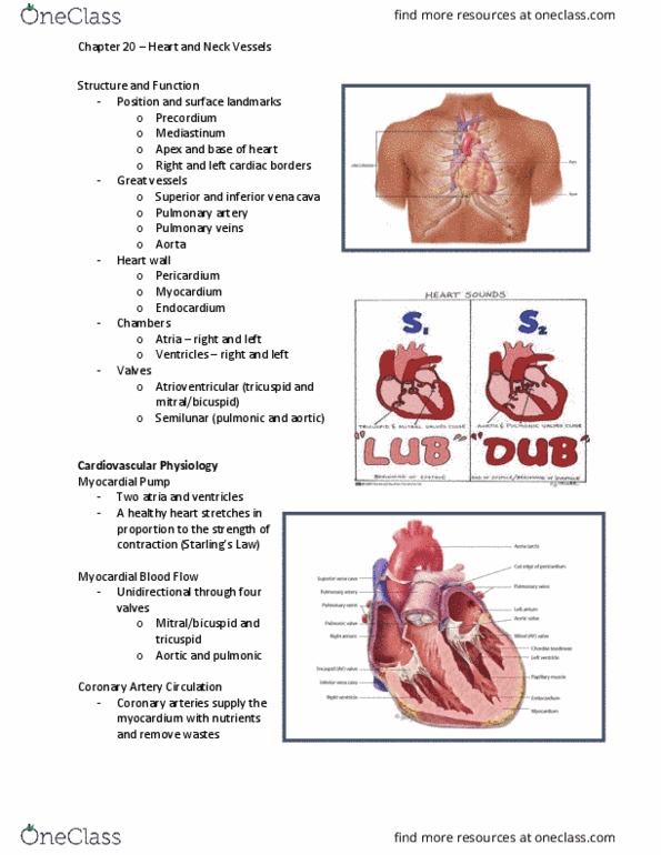
Hollow cone shaped muscular organ, size of fist. Located left of the midline, mediastinal cavity of the thorax between 2 nd and 5th intercostal space. Cardiac output- volume of blood ejected from each ventricle in one minute. Stroke volume- volume of blood ejected with each beat. Preload- the load that stretches the cardiac muscles before contraction. Myocardial contractility: ability of cardiac muscle when given a load, to contract or shorten. Afterload: degree of vascular resistance to ventricular contraction. Four chambers: right &left atria, right & left ventricle. Superior vena cava, right atrium, tricuspid valve, right ventricle, pulmonary valve, pulmonary artery, lungs, pulmonary vein, left atrium, bicuspid (mitral) valve, left ventricle, aortic valve, aorta. Sa node (60-100), av node (40-60), bundle of his (20-40), purkinje fibers. Hemodynamics- movement and pressure of the blood related to the electrical activity of the heart (heart sounds) Contraction of cardiac smooth muscle produced electrical activity. Monitors the electrical activity of the heart.