BIO 012 Lecture Notes - Lecture 18: Sliding Filament Theory, Skeletal Muscle, Cardiac Muscle

12 Jun 2018
School
Department
Course
Professor

Chapter 48 – Muscle Contraction
48.1 How Do Muscles Contract?
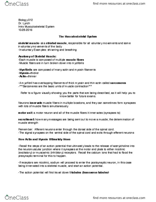
Three types of vertebrate muscle
- Skeletal Muscle – voluntary movements like running, and involuntary movements like breathing
or shivering
- Cardiac muscle – beating of the heart
- Smooth muscle -creates movement in many hollow internal organs, such as the digestive
system, bladder, and blood vessels, and is under the control of the autonomic (involuntary)
nervous system.
Muscle Structure
- Connections to bone
- Origins → less moveable and it anchors the muscle
- Insertion → ore oeale; tedo: oetie tissue o hih usle pulls
- Antagonist Muscles
o Flexor – decrease angle of joint
o Extensor – increase angle of joint
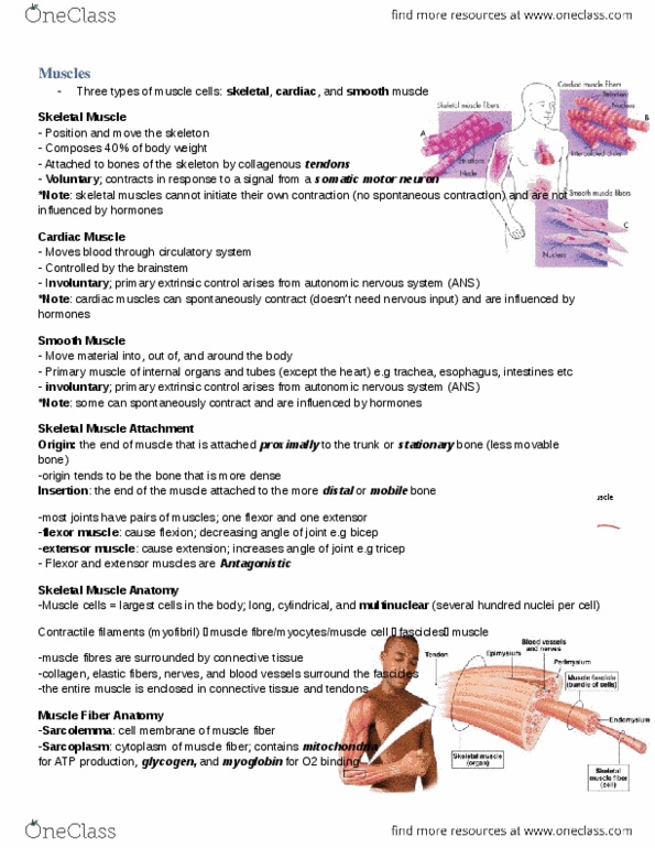
Skeletal Muscle
Skeletal muscles consist of bundles of muscle fibers which are large cells that have many nuclei. Skeletal
muscles also have many myofibrils which are bundles of actin and myosin filaments that form an
overlapping arrangement called sarcomeres which give the muscles their striated appearance. The
changes in the banding patterns of sarcomeres led to the sliding filament model of muscle contraction.
Skeletal muscle is called striated muscle because of its striped appearance. Skeletal muscle cells called
muscle fibers, are large and have many nuclei which form in the development through the fusion of many
individual embryonic muscle cells called myoblasts.
find more resources at oneclass.com
find more resources at oneclass.com

Point of contraction is to bring the Z-lines together of the sarcomere and the muscle contracts
- Myosin hooks unto the Z-lines and pulls both sides of the sarcomere together.
- The Z-lines are the vertical ends of the sarcomere
- Actin is the horizontal ends of the sarcomere
When the muscle is relaxed, the myosin is extended and only overlaps with actin at one small end part
- When the only thing in the middle is myosin, that zone is called the H zone
o With more overlap, the H zone decreases
- When there is only actin on the end, that zone is called the I band
o With more overlap, there I band decreases
- When the entire length of myosin overlaps with the actin, the area is called the A band
find more resources at oneclass.com
find more resources at oneclass.com

Muscle contraction is due to the interaction between the contractile proteins actin and myosin.
- One muscle cell has many myofibrils and therefore many sarcomeres
- Within muscle cells, actin and myosin molecules are organized into filaments. When muscle
contraction is triggered, the actin and myosin filaments slide past each other in a telescoping fashion.
- Each muscle fiber is packed with myofibrils → sarcomeres → bundles of actin and myosin
Troponin can push tropomyosin off the binding sites so that myosin can attach but this can only be done once
calcium binds to the troponin.
Conformational change – functional change in shape of a protein [Key word: function: the change of shape
has a function]
Regulation of Contraction (in the sarcomere) PROCESS
Relaxation
- Myosin heads not attached
- Tropomyosin (around actin) blocks the binding sites from letting myosin heads attach (little spots that
fit only a certain protein) on actin
- Troponin is attached to tropomyosin. It does not move without Calcium.
Contraction
- Calcium binds to troponin which moves the tropomyosin
- Tropomyosin now must move and uncover the myosin binding sites on the actin.
- Cross bridges form when myosin heads bind to actin and the heads begin to pull them
o This causes a conformational change
find more resources at oneclass.com
find more resources at oneclass.com
Document Summary
Skeletal muscle voluntary movements like running, and involuntary movements like breathing or shivering. Cardiac muscle beating of the heart. Smooth muscle -creates movement in many hollow internal organs, such as the digestive system, bladder, and blood vessels, and is under the control of the autonomic (involuntary) nervous system. Origins less moveable and it anchors the muscle. Insertion (cid:373)ore (cid:373)o(cid:448)ea(cid:271)le; te(cid:374)do(cid:374): (cid:272)o(cid:374)(cid:374)e(cid:272)ti(cid:448)e tissue o(cid:374) (cid:449)hi(cid:272)h (cid:373)us(cid:272)le (cid:862)pulls(cid:863) Antagonist muscles: flexor decrease angle of joint, extensor increase angle of joint. Skeletal muscles consist of bundles of muscle fibers which are large cells that have many nuclei. Skeletal muscles also have many myofibrils which are bundles of actin and myosin filaments that form an overlapping arrangement called sarcomeres which give the muscles their striated appearance. The changes in the banding patterns of sarcomeres led to the sliding filament model of muscle contraction. Skeletal muscle is called striated muscle because of its striped appearance.