BSC 1086C Lecture Notes - Lecture 7: Alveolar Cells, Pulmonary Vein, Bronchial Artery

Document Summary
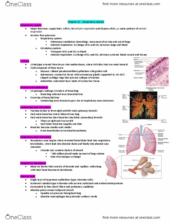
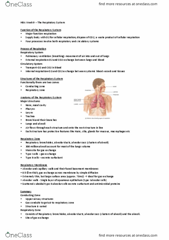
Respiratory bronchioles, alveolar ducts, alveolar sacs (cluster of alveoli: ~300 million alveoli account for most of the lungs" volume and are the main site for gas exchange. Respiratory membrane: ~ 0. 5-um-thick air-blood barrier: alveolar and capillary walls and their fused basement membranes, alveolar walls- single layer of squamous epithelium (type i cells, scattered type ii cuboidal cells secrete surfactant and antimicrobial proteins. Contain open pores that: connect adjacent alveoli, allow air pressure throughout the lung to be equalized. House alveolar macrophages that keep alveolar surfaces sterile. Right lung has 3 lobes, left lung 2: root: site of vascular and bronchial attachments. Costal surface: anterior, lateral, and posterior surfaces: apex: superior tip, base: inferior surface that rests on the diaphragm, hilum: on mediastinal surface; site for attachment of blood vessels, bronchi, lymphatic vessels, and nerves. Cardiac notch of left lung: concavity that accommodates the heart.