Biology 2382B Lecture Notes - Lecture 2: Differential Interference Contrast Microscopy, Antonie Van Leeuwenhoek, Optical Microscope

5 Feb 2018
School
Department
Course
Professor
Document Summary
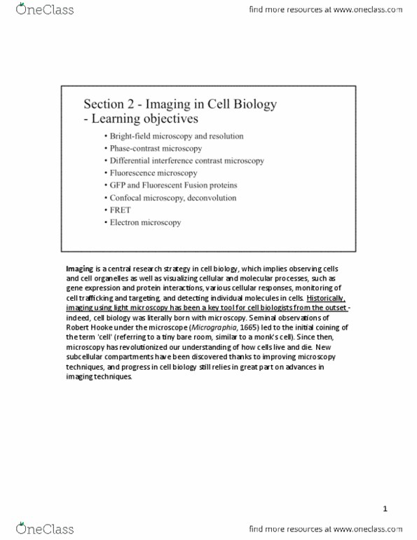
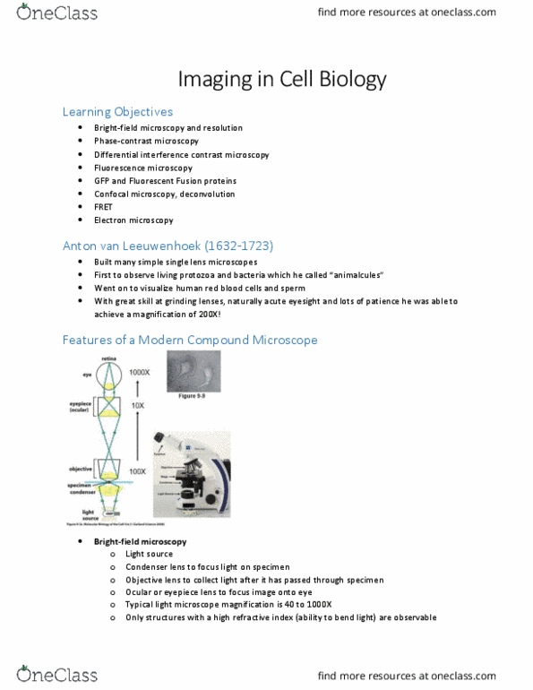
Animalcules : went on to visualize human red blood cells and sperm, he was able to achieve a magnification of 200x. Resolution of microscopes: resolution is the ability to distinguish between two very closely positioned objects as separate entities, a conventional light microscope usually can"t resolve objects/cellular features that are less than 0. 2 microns apart, smaller resolution is better. Wavelength spectrum used in microscopy: the visible spectrum is 400-700nm, light microscopy covers 300-700nm, getting into uv and x-rays, electron microscopy uses a very small wavelength, providing lower resolution. Phase contrast microscopy: phase contrast microscopy is used to examine live. How are fluorescence images obtained: the sample is illuminated from above, using a light source that incorporates from. Dual label fluorescence microscopy: when imaging dual label fluroesence-labelled structures, each structure is viewed individually with a specific microscope filter set for the fluorochrome, the images of the separate flurorochrome are then overlayed.