<From the desk of Michael Brennan>
B
[email protected]
Tue 9/19/2017 4:17 PM
To:
[email protected];
Here is the exam review for our next major lecture exam which will be on Monday. Notice that there are a few things missing, such as endochondral ossification. These will be added so just because it does not show up on this exam review doesn't mean you shouldn't study it. That being said, the details of each and every bone which I've gone over in lecture will be on the lab practical rather than the written lecture exam.
Anat 1, Exam 2. Spr 2016 LASC Name:____________________________________________________
1) A synarthrosis: a) is filled with synovial fluid. b) allow for a wide range of movement. c) includes the squamosal suture. d) is exemplified by the shoulder. e) all of the above.
2) Bursae: a) are located in the right atrium. b) are like elastomeric shock absorbers. c) are found in skeletal muscle fibers. d) form the sarcoplasmic reticulum. e) none of the above.
3) Irregular bones: a) can act like cams. b) can provide sites for muscle attachment. c) can protect soft-tissue organs. d) can act as levers. e) all of the above.
4) Condyles: a) are smooth articular surfaces. b) allow blood vessels to enter the bones. c) occur beneath the nail body after an injury. d) are produced by infected sebaceous glands. e) none of the above.
5) A head: a) is where you will find hyaline cartilage. b) is a smooth articular surface. c) is where a bone meshes with another bone. d) all of the above. e) none of the above.
6) Which of the following is the proper sequence of things following a bone fracture: a) hematoma, fibrous callus, bony callus, remodeling. b) hematoma, bony callus, fibrous callus, remodeling. c) hematoma, remodeling, fibrous callus, bony callus. d) hematoma, remodeling, bony callus, fibrous callus. e) fibrous callus, bony callus, remodeling, hematoma.
7) A diaphysis: a) contains yellow bone marrow. b) contain a lot of spongy bone tissue. c) occurs at the ends of long bones. d) all of the above. e) none of the above.
8) Short bones: a) protect underlying soft tissue organs. b) include the ribs. c) include the cranial bones of the skull. d) transfer mechanical forces. e) all of the above.
9) Fibrous joints: a) contain cruciate ligaments. b) include the pubic symphysis. c) are typically found in third class levering systems. d) include the lambdoid suture. e) all of the above.
10) Which of the following doesnât belong? a) tibiofibular joint. b) dentino-alveolar joint.
c) intervertebral disc. d) radioulnar articulation. e) cartilage.
11) Which of the following doesnât belong? a) atlanto-occipital joint. b) humero-ulnar joint. c) frontal suture d) acromio-humeral joint. e) tibiotarsal joint.
12) Hyaluronic acid: a) forms the extracellular matrix. b) is found in fibrous joints. c) keeps cartilage soft and pliable. d) is found in synostoses. e) none of the above.
13) First class levers: a) include the tibio-tarsal joint. b) have the effort between the fulcrum and the load. c) are like a teeter-totter. d) all of the above. e) none of the above.
14) Synchondroses: a) are typically synovial joints. b) allow for very limited movement. c) include sutures and gomphoses. d) are encased in synovial capsules. e) none of the above.
15) Parallel muscles: a) have long excursions. b) include the abdominal muscles. c) have excellent endurance. d) have low strength. e) all of the above.
16) Sphincteral muscles: a) enclose openings or passageways. b) are used when you talk.
c) surround the eyes. d) include the orbicularis oculi. e) all of the above.
17) Pennate muscle architecture: a) is typified by the pectoralis major. b) have high endurance.
c) generates high strength. d) has a longer excursion. e) all of the above
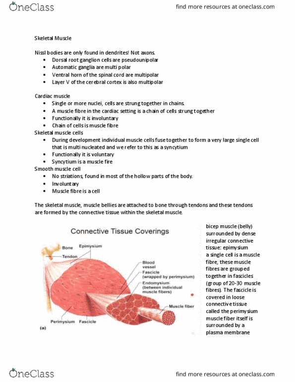
18) Endomysium: a) consists of dense fibrous connective tissue. b) surrounds each fasciculus.
c) surrounds bundles of muscle cells. d) all of the above. e) none of the above.
19) Cartilage: a) heals quickly if it is damaged. b) forms the lining of blood vessels. c) is found on the articular surfaces of bones. d) is a type of epithelial tissue. e) none of the above.
20) A tuberosity: a) is a hollow opening. b) is a shallow smooth articular surface. c) is a raised area that adjoins a condyle. d) is a larger outwardly curved articular surface. e) is a raised, rough-textured area.
21) Which of these doesnât belong? a) tuberosity b) trochanter c) condyle d) tubercle e) lunula
22) Epimysium: a) wraps around the entire muscle. b) is made of dense fibrous connective tissue. c) is continuous with tendons. d) all of the above. e) none of the above.
23) Which of the following is more likely to be an insertion rather than an origin? a) ulna.
b) cervical vertebra. c) thoracic vertebra. d) rib. e) Ischium.
24) Which of the following is more likely to be an origin rather than an insertion? a) clavicle.
b) tibia. c) radius. d) ulna. e) metatarsal.
25) Myofibrils: a) are composed of many muscle fibers. b) are composed of many muscle cells.
c) are composed of many sarcomeres. d) form the endomysium. e) all of the above.
26) Myosin: a) helps keep muscle cells from overstretching. b) is a protein. c) joins thin filaments together. d) forms part of the z-disc. e) all of the above.
27) Sarcolemma: a) surrounds the entire muscle organ. b) surrounds the endomysium.
c) is the cell membrane of a muscle fiber. d) surrounds the epimysium. e) none of the above.
28) Thin filaments: a) are made of titin. b) are pulled on by the thick filaments during contraction.
c) create the molecular âpower-strokeâ. d) all of the above. e) none of the above.
29) A second class lever: a) is like a wheel barrow. b) is like a teeter-totter. c) describes the humeroulnar joint. d) describes the atlanto-occipital joint. e) none of the above.
30) A sarcomere: a) is the basic unit of muscle function. b) is made mostly of proteins. c) shortens during a contraction. d) delivers an âall or noneâ response. e) all of the above.
31) Fast glycolytic fibers: a) have excellent endurance. b) deliver lots of power.
c) are more reddish in color than oxidative fibers. d) none of the above. e) all of the above.
32) Which of the following doesnât belong? a) Vastus lateralus. b) Vastus intermedius. c) Vastus medialis. d) Rectus Femoris. e) Gluteus minimus.
33) Which of the following doesnât belong? a) Teres major. b) Teres minor. c) subscapularis.
d) infraspinatus. e) supraspinatus.
34) Tendon sheaths: a) are filled with synovial fluid. b) can be likened to brake and shifter cable sheathing on a bike. c) are found around the carpal bones. d) really make life unpleasant if they get inflamed. e) all of the above.
35) Epithelial tissues: a) function in movement. b) conduct electrical impulses. c) form a structure for attachment of other organs. d) form barriers and walls. e) all of the above.
36) Elastic connective tissue: a) is found in bone. b) lines the inside of the trachea. c)Is found in small blood vessels like capillaries. d) is found in large arteries. e) all of the above.
37) Cartilage: a) heals quickly if it is damaged. b) forms the lining of blood vessels. c) is found on the articular surfaces of bones. d) is a type of epithelial tissue. e) none of the above.
38) Dense regular connective tissue: a) is poorly vascularized. b) heals slowly if at all. c) forms tendons and ligaments. d) can look a lot like smooth muscle under the microscope. e) all of the above.
39) Transitional epithelium: a) lines the urinary bladder. b) lines the blood vessels. c) contains cilia. d) joins the epidermis to the dermis. e) none of the above.
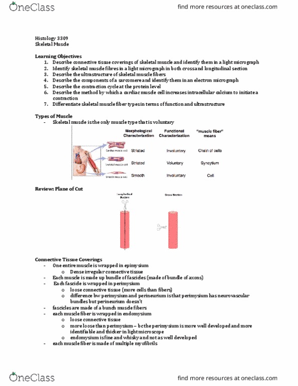
40) Skeletal muscle: a) responds to conscious thought. b) is found in the heart. c) is found in the walls of blood vessels and the GI tract. d) is composed of very small cells. e) all of the above.
For the following matching questions, each choice is used only once.
41) Fibroblast a) maintains bone tissue
42) Mast Cell b) secretes mucus
43) Chondrocyte c) responsible for inflammation response
44) Osteocyte d) makes collagen fibers
45) Goblet Cell e) maintains cartilage
For the following true-false questions, mark âAâ for true and âBâ for false.
46) Simple cuboidal epithelium is lines the trachea and Fallopian tubes/oviducts.
47) Elastic cartilage joins the ribs to the sternum.
48) An insertion is the more movable point of muscle attachment.
49) Tendons connect muscle to muscle or bone to bone.
50) Ligaments connect muscle to bone.
51) Transverse tubules allow nutrients to be dispersed throughout very large muscle cells.
52) Muscle twitch occurs when only a few muscle cells in the entire muscle are fully contracted.
53) Slow oxidative fibers have poor strength but excellent endurance.
54) Muscles can lengthen under their own power as well as contract.
55) Pottâs fracture is typical of skateboarders.
56) An incomplete fracture is a complete break through the entire bone.
57) Tetanus is the recruitment of additional fibers during muscle contraction.
58) Menisci are like elastomeric shock absorbers.
59) Diarthrotic joints allow for little to no movement.
60) A belly is the contractile portion of a muscle.
61) Synergistic muscles work with each other.
62) Muscle tissue, like your lab instructorâs evil twin before his morning coffee, is irritable.