Anatomy and Cell Biology 3309 Lecture Notes - Lecture 4: Loose Connective Tissue, Cardiac Muscle Cell, Micrograph

22 May 2018
School
Department
Professor

Histology 3309
Skeletal Muscle
Learning Objectives
1. Describe connective tissue coverings of skeletal muscle and identify them in a light micrograph
2. Identify skeletal muscle fibres in a light micrograph in both cross and longitudinal section
3. Describe the ultrastructure of skeletal muscle fibers
4. Describe the components of a sarcomere and identify them in an electron micrograph
5. Describe the contraction cycle at the protein level
6. Describe the method by which a cardiac muscle cell increases intracellular calcium to initiate a
contraction
7. Differentiate skeletal muscle fiber types in terms of function and ultrastructure
Types of Muscle
- Skeletal muscle is the only muscle type that is voluntary
Review: Plane of Cut
Connective Tissue Coverings
- One entire muscle is wrapped in epimysium
o Dense irregular connective tissue
- Each muscle is made up bundle of fascicles (made of bundle of axons)
- Each fascicle is wrapped in perimysium
o loose connective tissue (more cells than fibers)
o difference bw perimysium and perineurium is that perimysium has neurovascular
bundles but perineurium doesn’t
- fascicles are made of a bunch muscle fibers
- each muscle fiber is wrapped in endomysium
o loose connective tissue
o more loose than perimysium – bc the perimysium is more well developed and more
identifiable and thicker in light microscope
o endomysium is fine and whisky and not as well developed
- each muscle fiber is made of multiple myofibrils
find more resources at oneclass.com
find more resources at oneclass.com

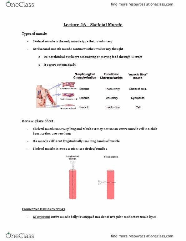
- this is a cross section of skeletal muscle
- MF = myofiber (aka muscle cell)
o Each myofiber is stippled (due to myofibrils)
- Endo= endomysium
o Loose connective tissue wrapping around each muscle cell (myofiber)
- Peri= perimysium
- Epi=epimysium
o Dense irregular connective tissue
o See collagen and fibroblasts
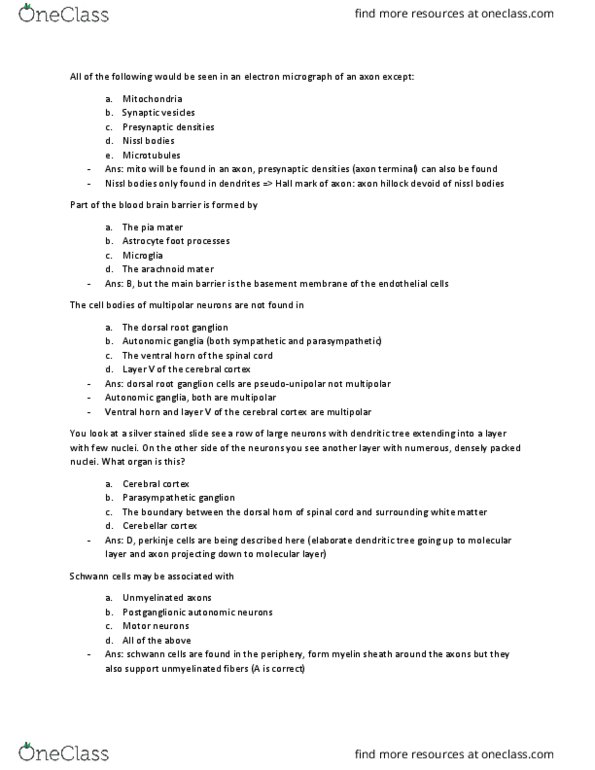
Skeletal Muscle Fiber (aka myofiber aka muscle cell) Ultrastructure
- Note: Sarcoplasmic reticulum not shown
- Myofibrils are made of the contractile units of the muscle (sarcomeres)
- Myofibrils are long tubes made of chains of sarcomere (made of contractile proteins of actin and
myosin)
o Striated appearance is due to overlap of contractile proteins (actin and myosin)
- It the overlap of contractile proteins within the myofibril that allows the muscle to contract as a
whole
- This pic shows 1 muscle cell
- Tons of mitochondria within the muscle cell bc skeletal muscle is very ATP dependent
- Muscle cells are multinucleated (nuclei located in periphery of cell)
- Sarcolemma (cytoplasm of cell) encompasses muscle cell
find more resources at oneclass.com
find more resources at oneclass.com

- Entire muscle cell is wrapped in sarcolemma
- Sarcolemma invaginates in bw all the myofibrils (called the T-tubule)
- Underneath the sarcolemma is the sarcoplasmic reticulum (dark blue) (like the ER)
- Skeletal muscle is a nonmembraenous network)
- As you get to the t-tubule, it becomes highly specialized and you get a triad made of…
o Continuation of sarcolemma
o 2 terminal cisternae (continuation of sarcoplasmic reticulum)
- on an EM image, if you see 3 little tubes, its going to be skeletal muscle (if you only see 2 tubes,
its going to be cardiac muscle)
- the triad is found at junction of A and I band
- triads are important for Ca2+ release in muscle contraction
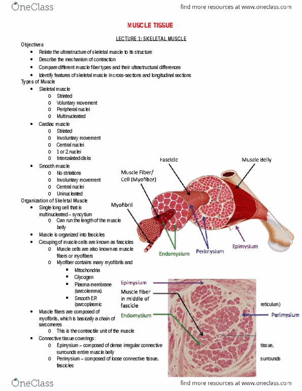
Muscle Fibers
- this is stained with H&E
- image on left = longitudinal section
o black outlined is one muscle cell
o parallel striated appearance due to overlap of myosin and actin (this is an identifying
feature)
o multinucleated with nuclei located in periphery
- image on right = cross section
o polygonal structure (bundles are all multiple shapes)
o multinucleated with nuclei located in periphery
o endomysium in bw all these bundles
o striation or stippled appearance in this cross section is due to myofibrils
find more resources at oneclass.com
find more resources at oneclass.com
Document Summary
Skeletal muscle is the only muscle type that is voluntary. One entire muscle is wrapped in epimysium: dense irregular connective tissue. Each muscle is made up bundle of fascicles (made of bundle of axons) Mf = myofiber (aka muscle cell: each myofiber is stippled (due to myofibrils) Endo= endomysium: loose connective tissue wrapping around each muscle cell (myofiber) Epi=epimysium: dense irregular connective tissue, see collagen and fibroblasts. Skeletal muscle fiber (aka myofiber aka muscle cell) ultrastructure. Myofibrils are made of the contractile units of the muscle (sarcomeres) Myofibrils are long tubes made of chains of sarcomere (made of contractile proteins of actin and myosin: striated appearance is due to overlap of contractile proteins (actin and myosin) It the overlap of contractile proteins within the myofibril that allows the muscle to contract as a whole. Tons of mitochondria within the muscle cell bc skeletal muscle is very atp dependent. Muscle cells are multinucleated (nuclei located in periphery of cell)