Anatomy and Cell Biology 3309 Lecture Notes - Intercalated Disc, Smooth Muscle Tissue, Cardiac Muscle Cell

12 Jul 2019
School
Department
Professor
Document Summary
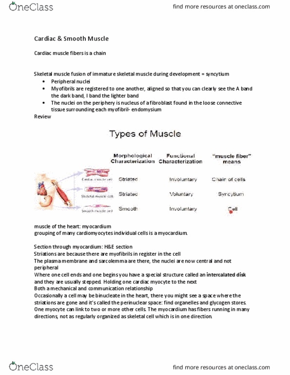
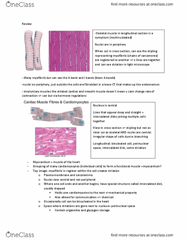
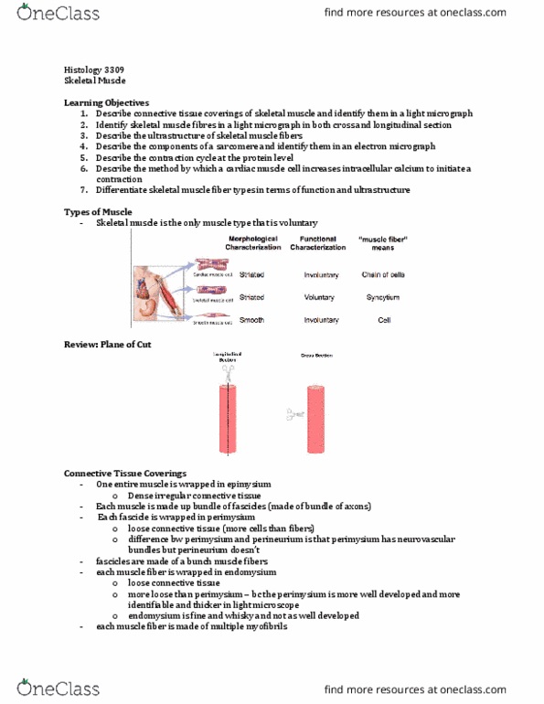
Cardiac muscle fibres: chain of cells (cardiomyocytes) attached to one another at intercalated discs. Cardiac & smooth muscle are both innervated by autonomic ns: a lot of smooth muscle in digestive tract, bladder, reproductive system. Cardiomyocytes come together to form 1 cardiac muscle fiber! (difference: skeletal muscle muscle cell = muscle fiber) Striations bc have sarcomeres: might not see full striations, bc of branches & plane of cut. Single, central nucleus; maybe rarely binucleate: organelles are at poles of nucleus (perinuclear space/cytoplasm, exception: lots of mitochondria found b/w myofibrils and run the length of the cell. Intercalated disks junction b/w adjacent cardiomyocytes: can be straight or jagged, abundant glycogen granules. Stippled appearance in x-section is from sarcomere arrangement (just like skeletal muscle) Cardiomyocyte ultrastructure: ultrastructural differences between cardiac and skeletal muscle cells, t tubules are larger in cardiac muscle and enter cell at z-disk (vs at the a-band/i-band junction in skeletal muscle)