Anatomy and Cell Biology 3309 Lecture Notes - Lecture 29: Efferent Ducts, Rete Testis, Seminiferous Tubule

22 May 2018
School
Department
Professor

Histology 3309
Male Ducts
Practice Question: ) form the blood testis barrier. ) secrete androgen binding protein. ) am an
epithelial cell. ) resorb residual bodies. Who am )?
a) Sertoli cell
b) Primary spermatocyte
c) Leydig cell
d) Spermatid
e) Spermatogonium
Learning Objectives
1. list in order the structures that spermatozoa pass through on their way from the seminiferous
tubule to the urethra.
2. describe the histological features of tubulusrectus, rete testis, efferent ductules, epididymis, and
vas deference
3. explain the functions of efferent ductules, epididymis, and vas deferens
4. list 3 glands associated with the male reproductive passages and name their functions.
5. draw a diagram of the general organization of the Prostate Gland
6. describe the histological organization of the erectile tissue in the penis
7. outline 4 physiological and cell biological steps the lead to penile erection
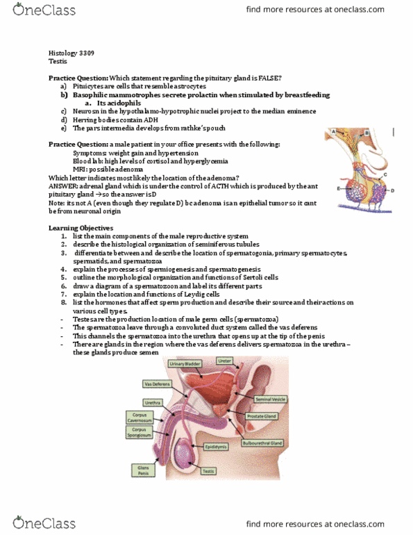
Sperm Transport and maturation
- sperm are collected in a meshwork called rete testis
- spermatozoa are collected into several tubules called ductuli efferentes (the ducts that carry
away from the testis)
- the ductuli efferentes combine into one highly coiled tube that has an elongated shape and lies
along the testis
- so the whole tube is called the epididymis but it starts from the head of the epididymis to the
body to the tail
- this tube is a single coiled tube that continues as a muscular epithelial lined tube called the vas
deferens you can see the tubules that line the testis on the side of the image enveloped in a
connective tissue
- the main job of the epididymis is the store spermatozoa
- they are then able to leave through the vas deferens
Fluid reabsorption occurs in efferent ductules
- rete testis is made of a simple cuboidal epithelium (cross section is shown it the blue image on
left)
- it forms an interconnected meshwork of these tubules that carry spermatozoa away from the
testis
- the rete testis carry the spermatozoa into the efferent ductules
find more resources at oneclass.com
find more resources at oneclass.com

o this epithelium is a tall pseudostratified epithelium
- the efferent ductules carry the spermatozoa into the highly coiled epididymis
o epithelium is very even (the outline is straight)
o the thickness of the epithelium is the same throughout
- rete testis is just a channel that transports spermatozoa along it doesn’t do anything else
- not only are spermatozoa transported, you also have a complex of testosterone and androgen
binding protein
- ABP sequesters testosterone and enriches it within the tubular fluid
- Testosterone is needed to maintain the function of these tubules
- Efferent ductules have an absorptive function – pump NaCl out of the lumen and create a high
concentration of NaCl just adjacent to the tubule
- Bc of the osmotic pressure, water is drawn out of the tubule
- So the spermatozoa are transported within a testicular fluid that is also produced by sertoli cells
bc they themselves are unable to swim away (they are still immature spermatozoa – haven’t
acquired motility yet)
- They acquire motility in the epididymis
- Note: another epithelium/organ that uses NaCl pumping as a way to concentrate something
(remove water from lumen) is the gallbladder
- Starting from the epididymis into the vas deferens, there is more and more smooth muscle
surrounding the tubule
o This is bc one of the functions of the vas deferens is to deliver the spermatozoa to the
outside world upon appropriate stimulation
o Ejaculation involves the contraction of smooth muscle that propels the spermatozoa to
the outside
Sperm transport from Testis to Rete Testis
- As the spermatozoa travel from the seminiferous tubule to the rete testis, there is a change in
the epithelium
- Recall: seminiferous tubule has spermatogenic cells surrounded by sertoli cells
- At one point, abruptly, spermatogenesis stops all of the sudden
o This is shown in the slide where it shows the seminiferous tubule opening into the rete
testis
o On the right side of the tubule, you only have sertoli cells (no more spermatogenic cells)
- The height of the epithelium (shown in green) changes to the cuboidal epithelium of the rete
testis
- Together, we have tight junctions (red bars) that form the blood testis barrier at the base of the
sertoli cells
find more resources at oneclass.com
find more resources at oneclass.com

- As the cell height changes, they tend to migrate to their normal epithelial position, further
towards the apical end
- Tubulus rectus
o Where you still have tall sertoli cells but no spermatogenesis going on
o Piece in bw the seminiferous epithelium and the rete testis epithelium that is straight
Ductuli Efferentes
- After the rete testis, it goes into the ductuli efferentes
- DISTINGUISHING FEATURE: spermatozoa in the middle
- The lumen of the ductuli efferentes has a saw tooth appearance
o Bc there are tall cells and stretches of shorter cells
- The epithelium of the ductuli efferentes is pseudostratified
- There are also basal cells stem cells that don’t reach the surface
- The taller cells often are ciliated
- Spermatozoa isn’t motilie yet so it has to be able to be transported from the seminiferous tubule
to the epididymis, embedded in this fluid →this is done by the cilia
- Principal cell:
o Shorter
o Not ciliated but they have stereocilia (modified microvilli) →this indicates an absorptive
function
o Principal cells is what removes NaCl and the water
o Abundant
- Ciliated cells:
o Keep the fluid in motion (help with motility)
find more resources at oneclass.com
find more resources at oneclass.com
Document Summary
Practice question: (cid:498)) form the blood testis barrier. ) secrete androgen binding protein. ) am an epithelial cell. ) resorb residual bodies. Who am )? (cid:499: sertoli cell, primary spermatocyte, leydig cell, spermatid, spermatogonium. Abp sequesters testosterone and enriches it within the tubular fluid. Testosterone is needed to maintain the function of these tubules. Not only are spermatozoa transported, you also have a complex of testosterone and androgen rete testis is just a channel that transports spermatozoa along (cid:523)it doesn"t do anything else(cid:524) Efferent ductules have an absorptive function pump nacl out of the lumen and create a high bc they themselves are unable to swim away (they are still immature spermatozoa haven"t. Bc of the osmotic pressure, water is drawn out of the tubule. So the spermatozoa are transported within a testicular fluid that is also produced by sertoli cells concentration of nacl just adjacent to the tubule acquired motility yet)