Anatomy and Cell Biology 3309 Lecture Notes - Lecture 43: Androgen-Binding Protein, Efferent Ducts, Rete Testis

2 May 2018
School
Department
Professor

Histology Lecture 15 – Semester 2
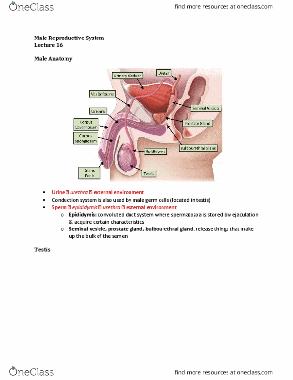
Male Reproductive System – MALE DUCTS
Questions
I form the blood-testis barrier, I secrete androgen binding protein, I am an epithelial cell. I resorb redirudal
bodies, which cell am I?
a. Sertoli cells
b. Primary spermatocyte
c. Leydig cell
d. Spermatid
e. Spermatoginium
Learning Objectives
1. List in order the structures that spermatozoa pass through on their way from the seminiferous
tubule to the urethra.
2. Describe the histological features of tubulusrectus, rete testis, efferent ductules, epididymis, and vas
deference.
3. Explain the functions of efferent ductules, epididymis, and vas deferens
4. List 3 glands associated with the male reproductive passages and name their functions.
5. Draw a diagram of the general organization of the Prostate Gland
6. Describe the histological organization of the erectile tissue in the penis
7. Outline 4 physiological and cell biological steps the lead to penile erection
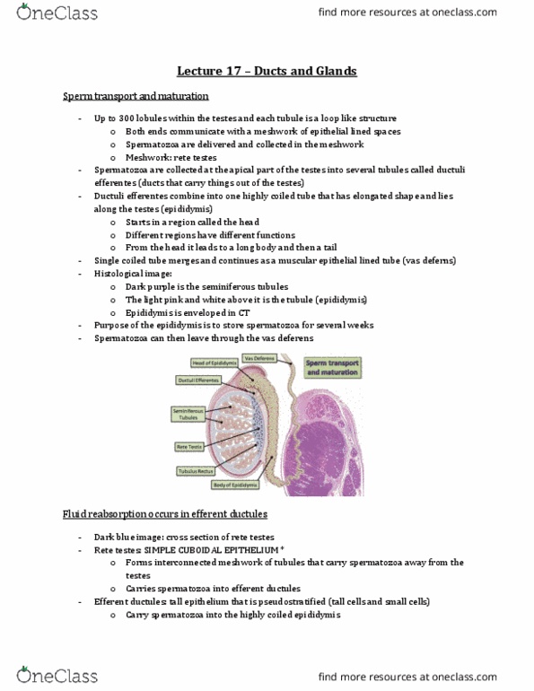
Sperm transport and maturation
- Up to 300 lobules in the testis
- Each tubule is a loop like structure – both ends communicate with a meshwork of epithelial lined
space
o Spermatozoa are being delivered and collected in this meshwork
o Since it is meshwork, name is Rete (net) Testis
- Spermatozoa are being collected at the apical part of the testis, into several tubules
o In humans, there are up to a dozen tubules = Ductuli Efferentes
▪ Ducts that carry OUT of the testes
- Ductuli Efferentes combine into one highly coiled tube, that has an elongated shape
o Lies along the testes
o = Epididymis
- In the epididymis, structurally there are structural and functional different within different regions
o Head, long body, tail
- Epidisymis is a single coiled tube that merges as a muscular epithelial lined tube = vas deferens
- In histological picture, see the seminiferous tubules
- Can make out these tubules
o Epididymis – highly coiled
o Lies alongside of the testes
find more resources at oneclass.com
find more resources at oneclass.com

o Enveloped into CT
- Main purpose of the epididymis = store spermatozoa
o After they are produced, they spend up to several weeks in the epididymis then they are
able to leave via the vas deferens
Fluid reabsorption occurs in Efferent Ductules
- Shows what the Rete Testes look like
o It is made up of simple cuboidal epithelium (Important)
o Forms interconnected meshwork of tubules that carries spermatozoa AWAY from the testes
- Rete Testis carries spermatozoa into the Efferent Ductules
o THE EPITHELIUM IS DIFFERENT – tall epithelium psuedostratified (has tall and smell cells)
o These ducts carry the spermatozoa into the epididymis (highly coiled)
- Epididymis is easy to identify because the epithelium is very even
o Outline is straight
o Thickness of the epithelium is the same throughout the length of the epididymis
- The rete testis is a channel that transports spermatozoa along
o Physiological function = conductive channel
o Not only spermatozoa being transported, but also a complex of testosterone and androgen
binding protein
▪ Androgen binding protein is important component that sequesters testosterone and
enriches it within the tubular fluid
▪ Androgen binding protein is produced by: Sertoli cells, under the influence of FSH
(hormone that induces the Sertoli cells to produce ABP)
- Testosterone is important to maintain the function of the tubules
o EX. Efferent ductile has absorptive function – there are cell types that pump NaCl out of the
lumen and create a high concentrate of NaCl just adjacent to the tubule
o Due to the osmotic pressure, water will be drawn out of the tubule
find more resources at oneclass.com
find more resources at oneclass.com

o Spermatozoa are transported within a testicular fluid that is also produced by Sertoli cells,
because they are unable to swim on their own (IMMATURE SPERMATOZOA THAT HAVE
NOT ACQUIRED MOTILITY)
- Motility is acquired in the epididymis
- Epithelium that uses NaCl pumping as a means of concentrating something or removing lumen from
something = gall bladder (does the same thing as the tubules in the testis)
- Purple line that thickens – starting from the epididymis into the vas deferens
o Along the length of the epididymis there will be more and more smooth muscle laid down
surrounding the tubule prominent in the vas deferens
- Function of the vas deferens (and a little bit of the epididymis): deliver the spermatozoa to the
outside world upon appropriate stimulation
o Ejaculation involves the contraction of smooth muscle that propels the spermatozoa to the
outside
Sperm transport from Testis to Rete Testis
- As the spermatozoa travel from the seminiferous tubule into the rete testis, there is a significant
change in the epithelial morphology
o Seminiferous tubule: spermatogenic cells surrounded by Sertoli cells
o At one point abruptly, the seminiferous tubule opens into the rete testis. Spermatogenesis all
of a sudden stop – ONLY HAVE SERTOLI CELLS LEFT (NO spermatogenic cells)
- The height of the epithelium changes to the cuboidal epithelium of the rete testis
- Tight junctions form the blood-testis barrier, are at the base of the Sertoli cells
o As the cell height changes, to migrate to their normal epithelium position, it is further to the
apical end
find more resources at oneclass.com
find more resources at oneclass.com
Document Summary
I form the blood-testis barrier, i secrete androgen binding protein, i am an epithelial cell. I resorb redirudal bodies, which cell am i: sertoli cells, primary spermatocyte, leydig cell, spermatid, spermatoginium. Up to 300 lobules in the testis. Each tubule is a loop like structure both ends communicate with a meshwork of epithelial lined space: spermatozoa are being delivered and collected in this meshwork, since it is meshwork, name is rete (net) testis. Spermatozoa are being collected at the apical part of the testis, into several tubules. In humans, there are up to a dozen tubules = ductuli efferentes: ducts that carry out of the testes. Ductuli efferentes combine into one highly coiled tube, that has an elongated shape: lies along the testes, = epididymis. In the epididymis, structurally there are structural and functional different within different regions: head, long body, tail. Epidisymis is a single coiled tube that merges as a muscular epithelial lined tube = vas deferens.