Anatomy and Cell Biology 3309 Lecture Notes - Lecture 3: Pancreas, Wisdom Tooth, Permanent Teeth

15 May 2018
School
Department
Professor

Lecture 3 – Teeth
Digestive system
1. Teeth
2. Tongue, salivary glands
3. Esophagus, stomach
4. Small and large intestine
5. Liver
6. Gall bladder, exocrine pancreas
- Teeth → epithelial lined tube
o There are glands on the outside of the tubular GI tract that are involved in digestion
(liver, pancreas, gallbladder)
Dentition
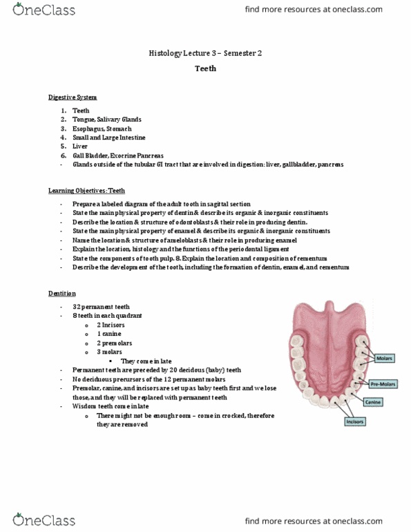
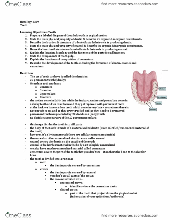
- Dentition: set of teeth we have
- 32 permanent teeth (ideally under normal circumstances)
- 8 teeth in each quadrant
o 2 incisors
o 1 canine
o 2 premolars
o 3 molars
o (5 molars)
- 3 molars come in late vs. the premolars, canine and incisors are set up as baby teeth first
and we lose them (they are replaced by permanent teeth)
o Wisdom teeth come in late and sometimes there is not enough room and they do not
come in straight
- Permanent teeth are preceded by 20 deciduous (baby) teeth
o Molas not replaced
- No deciduous precursors of the 12 permanent molars
find more resources at oneclass.com
find more resources at oneclass.com

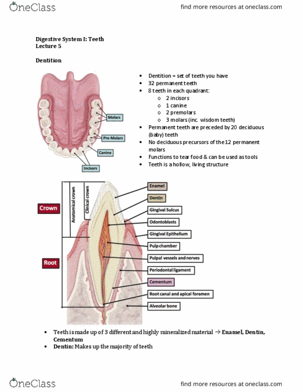
- IMPORTANT PICTURE – divides the tooth into different parts
- Dentin: body of the tooth is made of this material
o Goes all the way through the tooth and is the main calcified or mineralized hard
material of a tooth
o Dentin is a living material = there are cellular components inside (like osteocytes in
bone)
▪ If you knock your teeth out you can actually feel it – there is a signal
transduction going on
- Enamel: on top of the tooth
o Other mineralized structure
o Covers dentin on the outside of the tooth
o The hardest material in the body – HIGHLY MINERALIZED
- Cementum:
o Covers the part of the tooth that you do not see
o Anchors the tooth to the alveolar bone
o (tooth is anchored in a socket of alveolar bone)
o Another mineralized material
- Clinicians divide the tooth up into two regions:
o Root: covered by cementum
▪ Dentin part of the tooth that is part of the root is covered by cementum
o Crown: covered by enamel
▪ Do not see all parts of the crown
- Crown is divided into:
o Anatomical crown: identifies where the cementum starts
▪ Cementum covers the dentin on the root
▪ Crown: enamel
▪ Root: cementum
▪ In the root, the enamel covers the dentin on the crown
o Clinical crown: part of the tooth that projects from the gingivial sulcus (indentation
of epithelium/epidermis)
▪ Where problems arise because food/bacteria can get stuck = tooth decay
▪ An important clinical region
- Pulp: Tooth is hollow and has a chamber that is filled with loose CT
o All kinds of nerves and blood vessels come in to nourish the tooth
o Tooth is living entity and the cells that produce dentin line the pulp chamber and
they need nutrition/oxygen/waste removal
- Peridontal ligament (DCT): connection between the cementum and the alveolar bone
o Tooth must be anchored relatively firmly to the alveolar bone
o Ligament must be able to regenerate
o Leaves room for movement – do not really feel that teeth are wiggling, but they do
to a certain extent
o Peridontsist look closely at this region because if bacteria come in and start eating
away at the ligament, you can lose your teeth
find more resources at oneclass.com
find more resources at oneclass.com

o Orthodontists use this to move teeth – put pressure on the tooth to rearrange the
periodontal ligament in such a way that it can straighten the tooth
Development of teeth – how teeth are made
1. Bud stage
- Epithelium that lines the oral cavity starts to form growth into the underlying mesenchyme
o Mesenchyme: embryonic loose CT
- What induces the formation of a tooth bud is the accumulation of neural crest cells
o Accumulation of nuclei are seen under the tooth bud
o Neural crest cells migrate underneath the oral epithelium and locally induce the
epithelium to grow into the mesenchyme
- Cranial neural crest is important because cells have a very important function in forming
part of the tooth
- Neuroectodermal cells induce the overlying epithelial cells to proliferate and form an
invaginating tooth bud
find more resources at oneclass.com
find more resources at oneclass.com
Document Summary
Digestive system: teeth, tongue, salivary glands, esophagus, stomach, small and large intestine, liver, gall bladder, exocrine pancreas. Teeth epithelial lined tube: there are glands on the outside of the tubular gi tract that are involved in digestion (liver, pancreas, gallbladder) 32 permanent teeth (ideally under normal circumstances) 8 teeth in each quadrant: 2 incisors, 1 canine, 2 premolars, 3 molars, (5 molars) Permanent teeth are preceded by 20 deciduous (baby) teeth: molas not replaced. No deciduous precursors of the 12 permanent molars. Important picture divides the tooth into different parts. If you knock your teeth out you can actually feel it there is a signal transduction going on. Crown is divided into: anatomical crown: identifies where the cementum starts, cementum covers the dentin on the root, crown: enamel, root: cementum. Development of teeth how teeth are made: bud stage. Epithelium that lines the oral cavity starts to form growth into the underlying mesenchyme: mesenchyme: embryonic loose ct.