Anatomy and Cell Biology 3309 Lecture Notes - Lecture 39: Periodontal Fiber, Human Tooth Development, Cementum

2 May 2018
School
Department
Professor

Histology Lecture 3 – Semester 2
Teeth
Digestive System
1. Teeth
2. Tongue, Salivary Glands
3. Esophagus, Stomach
4. Small and Large Intestine
5. Liver
6. Gall Bladder, Exocrine Pancreas
- Glands outside of the tubular GI tract that are involved in digestion: liver, gallbladder, pancreas
Learning Objectives: Teeth
- Prepare a labeled diagram of the adult tooth in sagittal section
- State the main physical property of dentin & describe its organic & inorganic constituents
- Describe the location & structure of odontoblasts & their role in producing dentin.
- State the main physical property of enamel & describe its organic & inorganic constituents
- Name the location & structure of ameloblasts & their role in producing enamel
- Explain the location, histology and the functions of the periodontal ligament
- State the components of tooth pulp. 8. Explain the location and composition of cementum
- Describe the development of the tooth, including the formation of dentin, enamel, and cementum
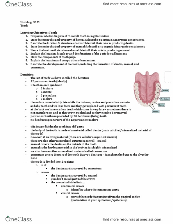
Dentition
- 32 permanent teeth
- 8 teeth in each quadrant
o 2 Incisors
o 1 canine
o 2 premolars
o 3 molars
▪ They come in late
- Permanent teeth are preceded by 20 decidous (baby) teeth
- No deciduous precursors of the 12 permanent molars
- Premolar, canine, and incisors are set up as baby teeth first and we lose
those, and they will be replaced with permanent teeth
- Wisdom teeth come in late
o There might not be enough room – come in crocked, therefore
they are removed
find more resources at oneclass.com
find more resources at oneclass.com

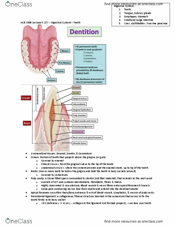
KNOW THIS SLIDE***
- Body of the tooth is made up of dentin
o Main calcified or mineralized hard material of the tooth
o It is a living material CELLULAR COMPONENTS INSIDE (similar to osteocyte in bone)
- Enamel covers the dentin on the OUTSIDE of the tooth
o HARDEST material in the body; highly mineralized
- Cementum covers the part of the tooth that you do not see (it anchors the tooth to the alveolar
bone)
o Tooth is anchored in a socket of alveolar bone
- There are two regions of the tooth:
o Root: covered by cementum
▪ Dentin part of the tooth that is part of the root is covered by cementum
▪ Part of the tooth that projects above the gingiva (gum)
o Crown: covered by enamel
▪ There are two parts:
• Anatomical crown: identifies where the cementum starts
o Cementum covers the dentin on the crown
o In the root, the enamel covers the dentin on the crown
• Clinical crown: part of the tooth that projects from the gingival sulcus
(indentation of your epidermis)
o Problem arises because food gets stuck
o Bacteria invades and have tooth decay
▪ You do not see all parts of the crown
▪ Portion of tooth that projects above the gingiva (or gum)
- The tooth is hollow – pulp chamber
o Has chamber that is filled with loose connective tissue, pulp
▪ All kind of nerves and blood vessels come in to nourish the tooth
▪ Tooth is a living entity
▪ Cells producing dentin are lining the pulp chamber, and they need nutrition, oxygen,
etc.
o Important part of the tooth
o Covered by dentin & extends to the root canal (apex of the root)
- Tooth has to anchored relatively firmly to the alveolar bone. There is a connection between the
centum and alveolar bone = periodontal ligament
o It is important for the ligament to be able to regenerate
▪ It is dense CT
▪ Leaves a little bit of room for movement
• The teeth wiggle to a certain extent
▪ If bacteria come and start eating away at the periodontal ligament, you can lose your
teeth
o Orthodontists use this to straighten teeth; put pressure on the tooth gently, and that
rearranges the periodontal ligament is such a way that it straightens the tooth
- Periodontal ligament = collagenous, fibrous structure inserted in the cementum that serves to fix the
tooth firmly in its bony socket
find more resources at oneclass.com
find more resources at oneclass.com

- The tooth is made of developmentally different parts of the body
1. Bud Stage
- Neuroectodermal cells induce the overlying epithelial cells to proliferate and form an invaginating
tooth bud
- Early on, the epithelium that lines the mouth (oral cavity) starts to form growth into the underlying
mesenchyme (Bulges into mesenchyme to form dental lamina in each jaw)
- Mesenchyme is the embryonic loose CT
- What induces the formation of the tooth bud is the accumulation of neurocrest cells
o Accumulation of nuclei underneath the tooth bud
o Neurocrest cells that migrate underneath the oral epithelium and locally induce the
epithelium to grow into the mesenchyme
- Cranial neurocrest cell ** is important function in forming part of the tooth
find more resources at oneclass.com
find more resources at oneclass.com
Document Summary
Digestive system: teeth, tongue, salivary glands, esophagus, stomach, small and large intestine, liver, gall bladder, exocrine pancreas. Glands outside of the tubular gi tract that are involved in digestion: liver, gallbladder, pancreas. State the main physical property of enamel & describe its organic & inorganic constituents. Prepare a labeled diagram of the adult tooth in sagittal section. State the main physical property of dentin & describe its organic & inorganic constituents. Describe the location & structure of odontoblasts & their role in producing dentin. Name the location & structure of ameloblasts & their role in producing enamel. Describe the development of the tooth, including the formation of dentin, enamel, and cementum. Explain the location, histology and the functions of the periodontal ligament. 8 teeth in each quadrant: 2 incisors, 1 canine, 2 premolars, 3 molars, they come in late. Permanent teeth are preceded by 20 decidous (baby) teeth. No deciduous precursors of the 12 permanent molars.