Anatomy and Cell Biology 3309 Lecture Notes - Lecture 47: Simple Columnar Epithelium, Intestinal Villus, Circular Folds

2 May 2018
School
Department
Professor

Histology Lecture 6 – Semester 2
Intestines
Learning Objectives
- Differentiate between the stomach, small intestine, and large intestine using histological markers
- Distinguish between plicae circulares, intestinal villi, and intestinal crypts
- List 4 ways in which the surface area of the intestinal tract is increased for absorption
o Plicae circulares = valves of Kerckring, semicircular folds of submucosa
o Intestinal villi = fingerlike projections of epithelium
o Microvilli = increase surface area of epithelial cells
o Glycocalyx = glycoproteins (enterokinases) projecting from microvilli
o Length of small intestine
- Describe the functions and locations of 4 cell types in the intestinal epithelium
- Outline the principles of lipid, protein, and carbohydrate absorption
- List histological features that distinguish the duodenum, ileum, and jejunum
- Explain the immunological significance of Peyer’s PatcheS
- Differentiate between cell renewal in the stomach, and in the small intestine
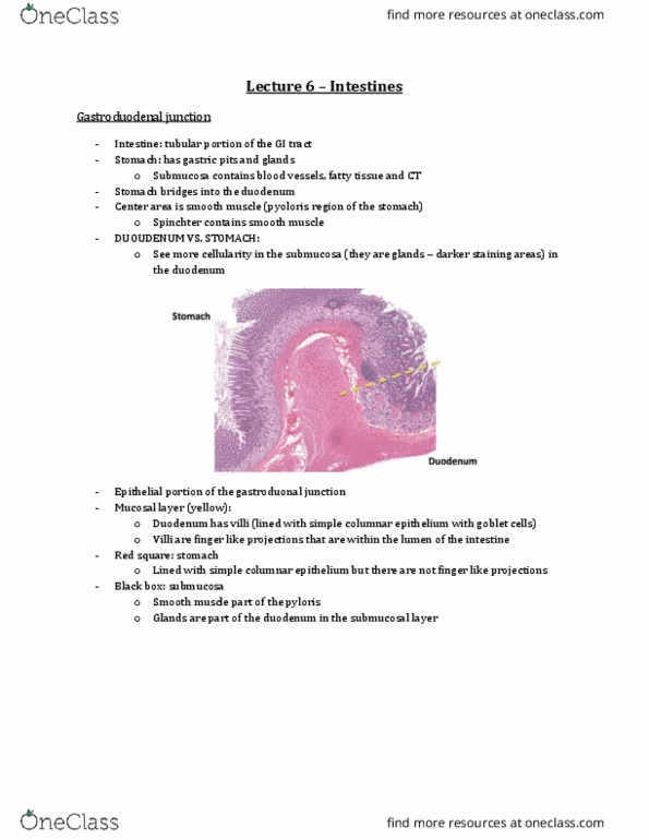
Gastroduodenal Junction
- See something more cellular in the submucosa = glands
o As you go from stomach to duodenum
More darker staining areas
- In the center there is smooth muscle = pylorus region of stomach
o Sphincter will have smooth muscle
find more resources at oneclass.com
find more resources at oneclass.com

- Mucosal layer
o Duodenum will have villi
▪ Lined with simple columnar epithelium with goblet cells
▪ Finger like projections into lumen of intestine
o Red square = stomach
▪ Lined with simple columnar epithelium but NO finger like projections
- Submucosa
o Black box = smooth muscle, part of pylorus
o Glands part of the duodenum in submucosal layer
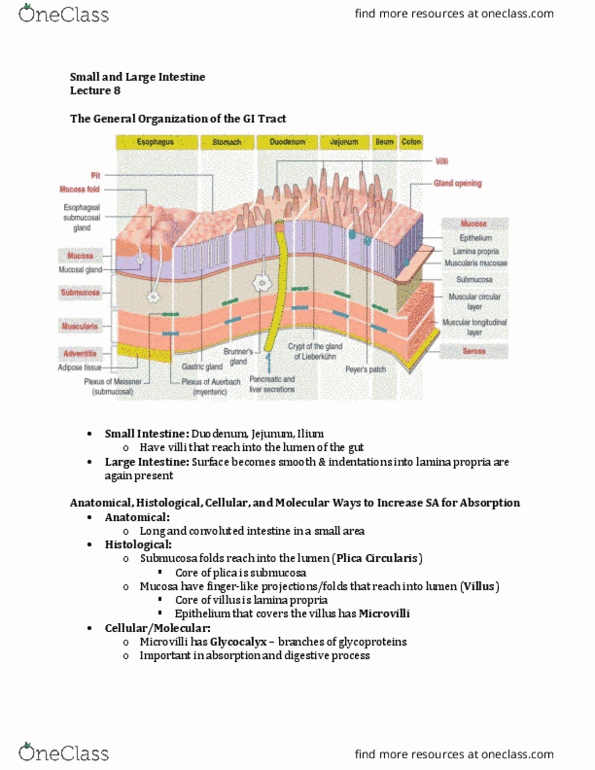
Main function of intestine = Absorption
- Small intestine = 20ft
- Large intestine = 5ft
- Increase SA to be able to reduce the length of the intestine that
has to do the same absorption
- Anatomical level we have, Plicae Circularis = folds of the
submucosa, protruding into the lumen of the intestine
- Tissue would look velvet. At the histological level we have villi
= lined with intestinal epithelium (simple columnar)
- Each cell of the epithelium has microvilli = tiny projections at
the top of the cell that increases absorption capacity
- On top of the microvilli, we have a layer that is secreted,
glycocalyx = glycoprotein, glycolipid based, increases the SA for
absorption
- Can only see it under EM because at the level of LM, we see a
blur of what is the microvilli and what is the glyocalyx
find more resources at oneclass.com
find more resources at oneclass.com

Villi
- Looking at the intestinal lumen, we will see the villi
- Villus covered by the epithelium
- At its core, there are lacteal = specialized lymphatic vessel that allows for transport of fatty
substances
o On each side, there is an arteriole and venule that allows for nutrient transport to systematic
circulation
- Look at the base of the villi, three are intestinal glands (Crypt of Lieberkuhn)
o Anything that is below the base of the villi is going to be your glands
find more resources at oneclass.com
find more resources at oneclass.com
Document Summary
Differentiate between the stomach, small intestine, and large intestine using histological markers. Distinguish between plicae circulares, intestinal villi, and intestinal crypts. List 4 ways in which the surface area of the intestinal tract is increased for absorption: plicae circulares = valves of kerckring, semicircular folds of submucosa. Intestinal villi = fingerlike projections of epithelium: microvilli = increase surface area of epithelial cells, glycocalyx = glycoproteins (enterokinases) projecting from microvilli, length of small intestine. Describe the functions and locations of 4 cell types in the intestinal epithelium. Outline the principles of lipid, protein, and carbohydrate absorption. List histological features that distinguish the duodenum, ileum, and jejunum. Differentiate between cell renewal in the stomach, and in the small intestine. See something more cellular in the submucosa = glands: as you go from stomach to duodenum. In the center there is smooth muscle = pylorus region of stomach: sphincter will have smooth muscle.