Anatomy and Cell Biology 3309 Lecture Notes - Lecture 6: Simple Columnar Epithelium, Lamina Propria, Intestinal Epithelium

15 May 2018
School
Department
Professor

Lecture 6 – Intestines
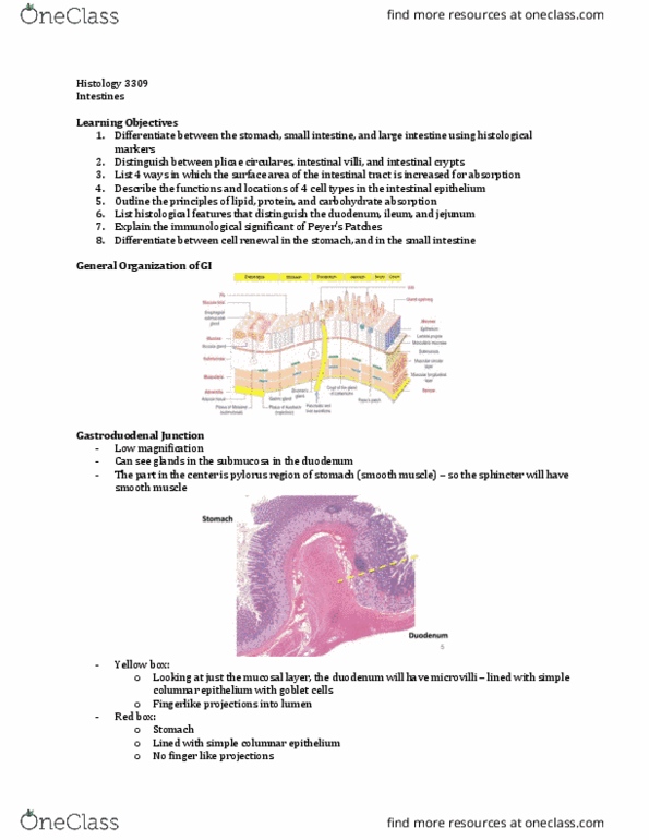
Gastroduodenal junction
- Intestine: tubular portion of the GI tract
- Stomach: has gastric pits and glands
o Submucosa contains blood vessels, fatty tissue and CT
- Stomach bridges into the duodenum
- Center area is smooth muscle (pyoloris region of the stomach)
o Spinchter contains smooth muscle
- DUOUDENUM VS. STOMACH:
o See more cellularity in the submucosa (they are glands – darker staining areas) in
the duodenum
- Epithelial portion of the gastroduonal junction
- Mucosal layer (yellow):
o Duodenum has villi (lined with simple columnar epithelium with goblet cells)
o Villi are finger like projections that are within the lumen of the intestine
- Red square: stomach
o Lined with simple columnar epithelium but there are not finger like projections
- Black box: submucosa
o Smooth muscle part of the pyloris
o Glands are part of the duodenum in the submucosal layer
find more resources at oneclass.com
find more resources at oneclass.com

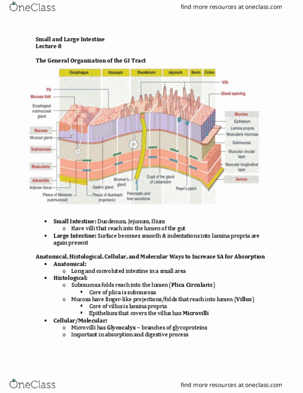
Main function of intestine = absorption
- Small intestine is 20 feet long and large intestine is 5 feet long
- Do not want small intestine to be any longer
o Body has mechanisms that increases the surface area to be able to reduce the actual
length of the intestine that has to do the absorption
- Increase surface area
- For efficacy purposes at the anatomical level, there are plicae circularis
- Plicae circularis are folds of the submucosa that can be seen protruding the lumen of the
intestine
o Tissue has a velvet appearance due to the villi
- Villi are lined by intestinal epithelium (simple columnar)
- Each cell of the epithelium has microvilli
o Microvilli: tiny projections at the top of the cell that also increase absorption
capacity
- On top of that, there is a glycocalyx layer that is secreted that is glycolipid/glycoprotein
based that also increases the surface area for absorption
o Glycocaylx is only seen in an EM – in an LM there is blur of what is the microvilli and
what is the glycocaylx
find more resources at oneclass.com
find more resources at oneclass.com

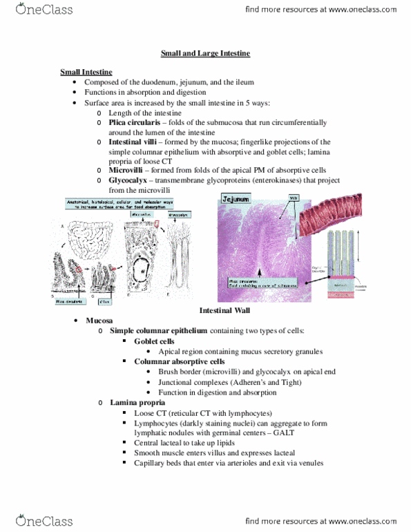
Villi
- Villi are in the intestinal lumen
- Each villus is lined by epithelium and at the core, there is a lacteral
- Lacteal: lymphatic vessel which allows for transport of fatty substances
o On each side, there is an arteriole and venule which allows for nutrient transport
into the systemic circulation
- At the base of the villi, there are intestinal glands (Crypt of Lieberkuhn)
o Anything below the villi are glands
Villi – longitudinal
- Lacteal is in the center
- Can see a lining around the lacteal (wall of the lymphatic vessel)
- There is some smooth muscle in the core of the villus
o Derived from muscularis mucosa
o Help contract the lacteal to move the lymph from the villus
- Epithelium sits on the lamina propria (contains fibroblasts, plasma cells, lymphocytes,
macrophages, etc)
find more resources at oneclass.com
find more resources at oneclass.com
Document Summary
Stomach: has gastric pits and glands: submucosa contains blood vessels, fatty tissue and ct. Center area is smooth muscle (pyoloris region of the stomach: spinchter contains smooth muscle. Duoudenum vs. stomach: see more cellularity in the submucosa (they are glands darker staining areas) in the duodenum. Mucosal layer (yellow): duodenum has villi (lined with simple columnar epithelium with goblet cells, villi are finger like projections that are within the lumen of the intestine. Red square: stomach: lined with simple columnar epithelium but there are not finger like projections. Black box: submucosa: smooth muscle part of the pyloris, glands are part of the duodenum in the submucosal layer. Small intestine is 20 feet long and large intestine is 5 feet long. Do not want small intestine to be any longer: body has mechanisms that increases the surface area to be able to reduce the actual length of the intestine that has to do the absorption.