BIO210Y5 Lecture 8: Heart - chp 19

17 Jan 2016
School
Department
Course
Professor
Document Summary
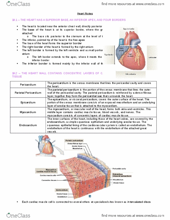
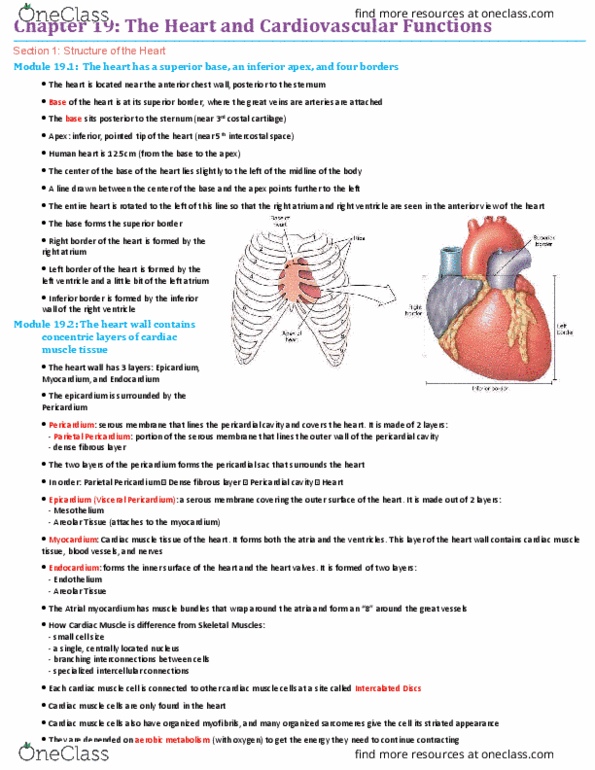
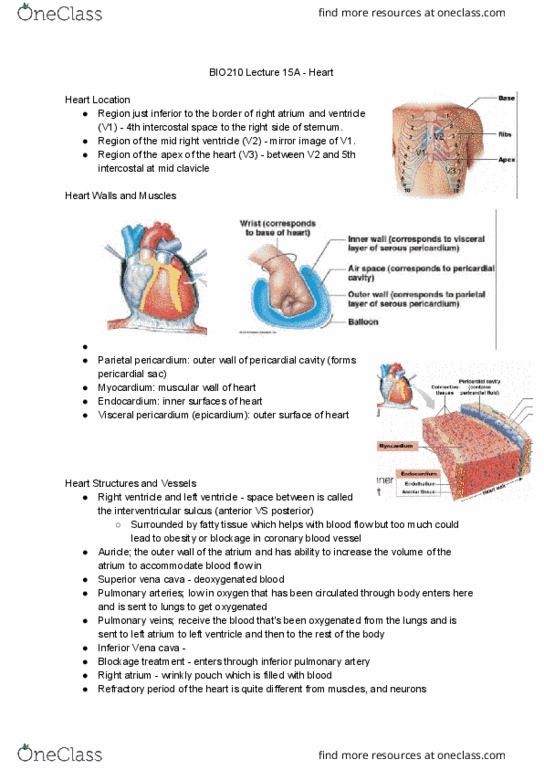
Myocardium: middle layer + muscular wall of the heart. Forms both atria + ventricles, contains cardiac muscle tissue, blood vessels & nerves. Endocardiu m: covers inners surfaces + valves of heart. Contains a squamous epithelial lining endothelium + areolar tissue. Pericardium: serous membrane, lines pericardial cavity, covers the heart. Parietal pericardium: portion of serous membrane that lines outer wall of pericardium cavity. Parietal pericardium + fibrous layer pericardial sac, surrounds the heart. Covers the outer portion of the heart. Fossa ovalis oval depression, remnant of foramen ovale, that closes at birth. Opening of right ventricle boarded by right atrioventricular valve (av) = tricuspid valve. Left atrioventricular valve=bicuspid valve=mitral valve permits the blood flow from left atrium in left ventricle, but prevents backflow during ventricular contraction. (same function as right av valve) If chordae tendineae are cut/papillary muscles are damaged regurgitation (backflow) of blood into atria occurs each time ventricles contract.