HSS 1100 Lecture Notes - Lecture 3: Hans Christian Gram, Gram Staining, Crystal Violet

26 Sep 2018
School
Department
Course
Professor
HSS 1100 verified notes
3/13View all
2
HSS 1100 Lecture Notes - Lecture 2: Complement Membrane Attack Complex, Innate Immune System, Vacuum Cleaner
3
HSS 1100 Lecture Notes - Lecture 3: Hans Christian Gram, Gram Staining, Crystal Violet
4
HSS 1100 Lecture Notes - Lecture 4: Lancefield Grouping, Streptococcal Pharyngitis, Opportunistic Infection
Document Summary
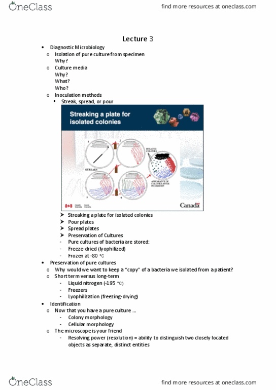
Because from the isolation you can make a diagnosis. Short term vs long term: liquid nitrogen (-195 c, freezers, lyophilization (freeze-drying) Now that you have a pure culture. : colony morphology, cellular morphology. The microscope is your friend: resolving power (resolution) = ability to distinguish two closely located objects as separate, distinct entities. Generally 3 steps: make a smear on inoculating colony, fix dried smear by heat fix, stain with desired dye. Simple stain: single dye normally used, all organisms same colour, size, shape, number, arrangement, etc. Differential stain: two or more dyes, differences between microorganisms or parts of cells, acid fast, gram. Gram positive: thick peptidoglycan, no 2nd cell wall, not able to produce and liberate endotoxin. Gram negative: thin peptidoglycan, can produce and liberate endotoxin. Shape of bacteria related to peptidoglycan layer. Gram negative usually thinner than gram positive. Antibodies tagged with dyes are common (immunofluorescence microscopy)