BIO 1140 Lecture Notes - Lecture 3: Spindle Apparatus, Cadherin, Centrosome

27 Feb 2017
School
Department
Course
Professor
Document Summary
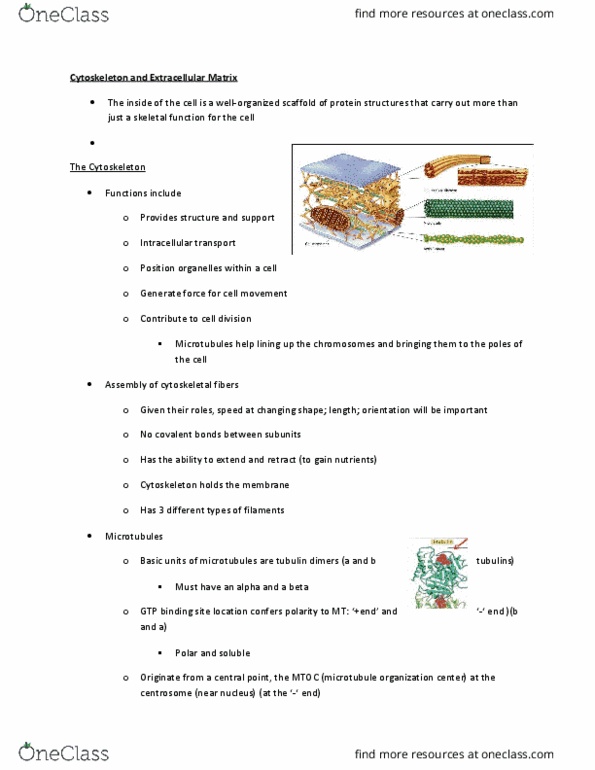
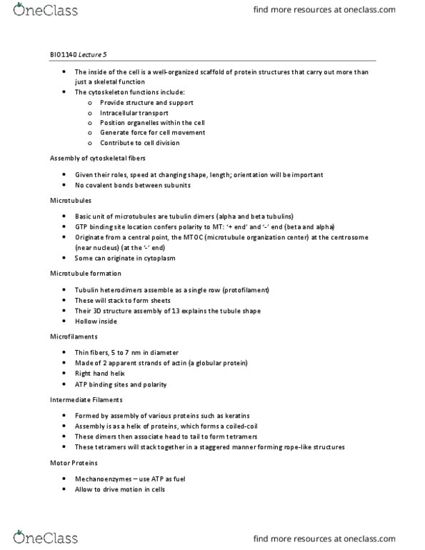
The inside of the cell is a well organized scaffold of protein structures that carry out more than just a skeletal function for the cell. The cytoskeleton: provides structure and support (the cell and organelles) Intracellular transport: position organelles within the cell, generate force for cell movement, contributes to cell division. Assembly of cytoskeletal fibers: given their roles, speed at changing shape, length; orientation will be important, no covalent bonds between subunits. Microtubule formation: tubulin heterodimers assemble as a single row (protofilament, these will stack to form sheets, their 3d structure assembly of 13 (1 every 28degrees) explains the tubule shape, hollow inside. Microfilaments: thin fibers, 5 to 7 nm in diameter, made of two apparent strands of actin (a globular protein, right hand helix, atp binding sites and polarity. Motor proteins: mechanoenzymes use atp a fuel, allow to drive motion in cells. Vesicles, organelles, pigments, membrane, motility and mobility, etc.