ANP 1105 Lecture Notes - Lecture 1: Systolic Geometry, Angina Pectoris, Aorta

11 Dec 2015
School
Department
Course
Professor
Document Summary
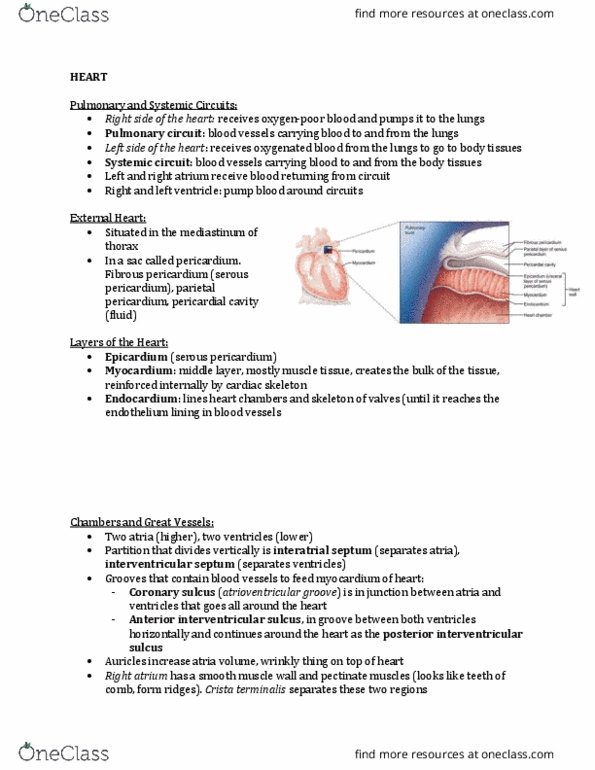
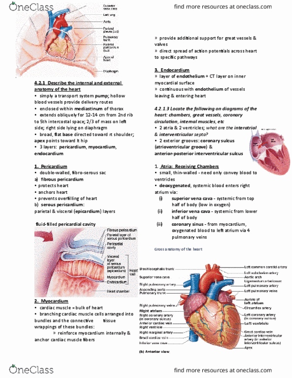
The heart is the size of your fist. It looks like a solid organ but it is just a hollow chamber inside. There are blood vessels running on the outside. The right ventricle is what you normally see, and the left ventricle is more in the posterior. Coranary sulcus: atrioventricular groove: anterior-poterior interventricular sulcus, important when it comes to the blood supply in the heart. Pulmonary trunk (artery) (deoxygenated), four pulmonary veins take oxygenated blood into the heart. Left atrium: oxygenated blood comes into the left atrium via pulmonary veins, pumps it into the left ventricle. Left and a right pulmonary trunk (goes into the left and the right lungs) The left ventricle has a thicker wall (myocardium) because it has to get the blood throughout the body and the right ventricle doesn"t because it just has to get blood into the lungs. The fossa ovalis: a depression; an oval; therefore an oval depression.