ANP 1105 Lecture Notes - Lecture 2: Posterior Interventricular Sulcus, Anterior Interventricular Sulcus, Interventricular Septum

6 Sep 2018
School
Department
Course
Professor
Document Summary
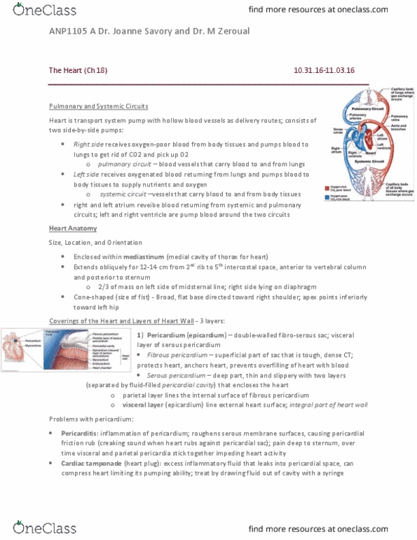
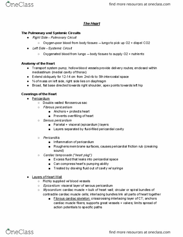
External heart: situated in the mediastinum of thorax. Fibrous pericardium (serous pericardium), parietal pericardium, pericardial cavity (fluid) Chambers and great vessels: two atria (higher), two ventricles (lower, partition that divides vertically is interatrial septum (separates atria), interventricular septum (separates ventricles, grooves that contain blood vessels to feed myocardium of heart: Coronary sulcus (atrioventricular groove) is in junction between atria and ventricles that goes all around the heart. Crista terminalis separates these two regions: right atrium, blood enters from 3 veins: Superior vena cava (returns blood from body regions superior to diaphragm) Inferior vena cava (returns blood from below diaphragm: left atrium is mostly smooth, pectinate is found in auricle. Coronary arteries: left and right coronary arteries arise from the base of the heart and circle around it in the coronary sulcus, they feed the myocardium but are in the epicardium (dip in the myocardium)