CAPS 390 Lecture Notes - Lecture 30: Common Hepatic Duct, Exocrine Gland, Immunostaining

18 Nov 2015
School
Course
Professor
Document Summary
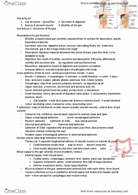
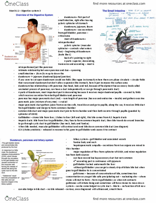
Lecture 30, gi -iii, nov 12, 2014 (dr. roskelley) Ch 18, pp 417-436 (note: you will not be tested on salivary glands in this course) Develop from evaginations of endodermal tube near the start of small intestine. Therefore, all three glands drain into the small intestine (pancreas, liver, and gall bladder) Beginning as an endodermal tube, gall bladder branches out as an out-pouching. Further from the out- pouching, the liver develops and forms and is joined by the common hepatic duct. Ventral part of pancreas develops with the out pouching, and joins with dorsal part of pancreas which develops opposite to the out pouching. All ducts empty at the sphincter of oddi into the small intestine. Pale/chromophobic cells with h & e staining (l. m. ) Two major cell types, discerned by immunostaining specific for peptide hormones/secretory product: Cells = secrete glucagon which increases the blood glucose. Cells = secrete insulin which decreases the blood glucose.