BLG 311 Lecture Notes - Lecture 2: Drug Delivery, Immunocytochemistry, Aquaporin

29 Dec 2017
School
Department
Course
Professor
Document Summary
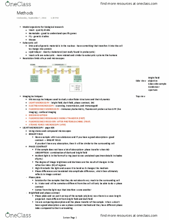
Types of light microscopy (bright field, dark field, phase-contrast, dic) Imaging tools needed to observe cells: light microscopy. Fluorescent proteins: gfp and others and related technology (dynamics, photoactivation, frap) Cells are too small for our eyes to resolve: A typical animal/human cell is around 10-100 micrometers. The human eye can only see/resolve particles that are >100 micrometers. Light rays are bent when entering and leaving different mediums (water, glass, and air) Objective and ocular lens magnify the image of the specimen by bending light. The condenser on the light microscope focuses light on the specimen. Resolution: resolution allows us to see the space between objects and able to see two separate molecules or particles rather than one giant big blurry blob. Detection: the detection is controlled by the exposure time (how long you are accepting light information) light microscopy. A: light microscopes magnify and increase the resolution of an image.