BCH 261 Lecture Notes - Lecture 8: Enthalpy, Streptococcus, Ribonuclease

7 Jun 2018
School
Department
Course
Professor

!
1!
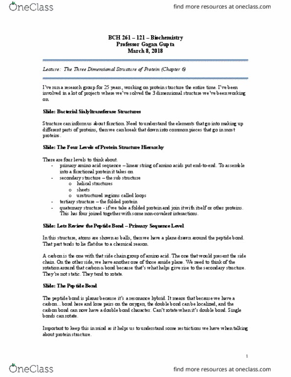
BCH 261 – 121 – Biochemistry
Professor Gagan Gupta
March 15, 2018
Mid-term: Average 67%
Lecture: Tertiary Structure
Slide: Ramachandran Plot
Biochemist studied protein structures solved by crystallography.
This plot is the sine angle on y-axis and Φ angle down here. Can see where all these things are
clustered.
Picture of secondary structure – two forms of ß sheets, parallel and anti-parallel. The Φ
side…bigger than 120. Α helices clustered down here, angles between 0 and 60. There are dots
elsewhere that are outliers from unusual structures.
Left handed helix – polypeptide left handed helix.
What you can get from this plot would be an allowed angle or secondary structure.
Slide: Steric Interactions Determine Peptide Conformation
If angles are too close together we get a steric clash at the top.
Slide: What Does the Angle…
Don’t have to remember the angles. The Φ side structures are bigger than in ß helix.
Slide: Classes of Tertiary Structures
- Fibrous proteins – your hair. Typically insoluable. Have a single secondary structure.
Skin and connective tissues.
- Globular proteins – globular in shape. Typically water-soluble. Enzymes. To carry out a
chemical reaction probably want some kind of cavity in the protein. Don’t need to carry
out any reactions. They are there for strength.
Slide: Fibrous Protein Structure
- keratin – hair, fingernails, feathers, scales
- fibroin – silk cocoons
- collagen – what holds your tissue together
!
2!
Slide: α-keratin
Two strands of keratin. Parallel arrangement. Take the long strand and turn it 90º, you’d be
looking down those two coils. There’s a super coil to this helix. Extra level of curve coming from
the amino acid composition.
Keratin has large hydrophobic residue repeats.
Hydrophobic residues on one side of helix for both members and that’s what helps the hair of
fibres stick together.
Coiled-coil structure.
Slide: Fibroin Fibers Emerge from Silk…
Another example of a fibrous protein.
Slide: Silk Fibroin: a ß sheet Protein
The orientation of the strands of the helix. Here we have the anti parallel arrangement and some
parallel arrangements. We have this mixture of these ß-sheets and they all stack on top of each
other. Look at the structure with the carbon backbone, we have an extended sheet and one sheet
sitting on top of another giving us a nice packed three-dimensional structure. Think about what
would that mean for the function or how this protein exists in nature. It will give that protein
some strength, but it’s flexible. It’s not rigid.
Slide: Questions for Next Class
(see slide)
Slide: Three Ways to Represent the 3D Structure: Human Ubiquitin
In the early days when people were trying to figure out what proteins looked like, we were
lacking the tools so ended up adapting methodology that had been used…beaming x-rays at a
crystal of a protein. Looking at reflected x-rays then coming up with a chemical representation.
Can do this remotely now. You end up with a 3D coordinate set we need to look at graphically.
We looked at these cartoon like structures where we’ve converted the real structure into a cartoon
structure. You can do it as a stick model and then that allows us, when we have a close up, to pay
attention to things like where hydrogen bonds would be, or how the atoms would interact.
Something you can’t get at when using this cartoon structure.
Slide: Solvent-Accessible Surface Models of Ubiquitin
Atom colouring, blue is nitrogen, red is oxygen, in this picture creating this carbon. You have
some idea of the atoms on the surface. Sometimes we want it to be monochrome so we can
emphasize there’s a cavity or opening here we might be interested in. You can calculate the
charge distribution on the surface of the protein with caveats – calculating surface potentials is
usually done in the gas phase, so these calculations don’t take into account water, so we take
them with a grain of salt. They are still useful guides to tell us there’s a positively charged patch
here in blue and negatively charged patch in red.
Document Summary
This plot is the sine angle on y-axis and angle down here. Can see where all these things are clustered. Picture of secondary structure two forms of sheets, parallel and anti-parallel. Helices clustered down here, angles between 0 and 60. There are dots elsewhere that are outliers from unusual structures. Left handed helix polypeptide left handed helix. What you can get from this plot would be an allowed angle or secondary structure. If angles are too close together we get a steric clash at the top. The side structures are bigger than in helix. To carry out a chemical reaction probably want some kind of cavity in the protein. Slide: fibrous protein structure keratin hair, fingernails, feathers, scales fibroin silk cocoons collagen what holds your tissue together. Take the long strand and turn it 90 , you"d be looking down those two coils. Extra level of curve coming from the amino acid composition.