BCH 261 Lecture Notes - Lecture 7: Beta Sheet, Polyproline Helix, Amphiphile

7 Jun 2018
School
Department
Course
Professor

!
1!
BCH 261 – 121 – Biochemistry
Professor Gagan Gupta
March 8, 2018
Lecture: The Three Dimensional Structure of Protein (Chapter 6)
I’ve run a research group for 25 years, working on protein structure the entire time. I’ve been
involved in a lot of projects where we’ve solved the 3 dimensional structure we’ve been working
on.
Slide: Bacterial Sialyltransferase Structures
Structure can inform us about function. Need to understand the elements that go into making up
different parts of proteins, then we can break that down into common pieces that go in most
proteins.
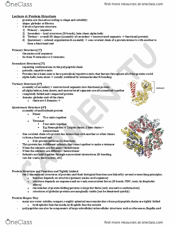
Slide: The Four Levels of Protein Structure Hierarchy
There are four levels to think about.
- primary amino acid sequence – linear string of amino acids put end-to-end. To assemble
into a functional protein it takes on
- secondary structure – the sub structure
o helical structures
o sheets
o unstructured regions called loops
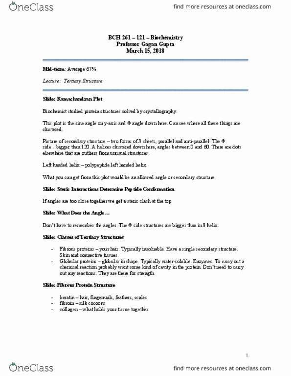
- tertiary structure – the folded protein
- quaternary structure - if we take a folded protein and join it with itself or other proteins.
This has four joined together with some non-covalent interactions.
Slide: Lets Review the Peptide Bond – Primary Sequence Level
In this structure, atoms are shown as balls, then we have a plane drawn around the peptide bond.
That part tends to lie flat due to a chemical reason.
Α carbon is the one with that side chain group of amino acid. The one that would present the side
chain. On the other side, we have another one of those amide plane. We need to think of the
rotation around that carbon α bond because that’s what helps give rise to the secondary structure.
They’re not static. They tend to rotate.
Slide: The Peptide Bond
The peptide bond is planar because it’s a resonance hybrid. It means that because we have a
carbon…bond here and lone pairs on the oxygen, the double bond can be localized, and the
carbon bond can now have a double bond character. Can’t rotate when it’s double bond. Single
bonds can rotate.
Important to keep this in mind as it helps us to understand some restrictions we have when talking
about protein structure.
!
2!
Slide: Components of a Protein
This is a protein used in the lab a lot. It binds to sugar molecules. This is a cartoon representation
of the three dimensional structure. I’ve coloured the secondary structure elements so you can
differentiate it.
Beta sheet is in magenta colour.
We like to think about the content of the protein is comprised of each of these secondary structure
elements? Are they all α-helical, beta sheet or mixed?
Slide: The α-helix
This molecule is constrained, it’s wrapped. If we put the side chains on then you can see how the
peptide bond fits in there. A key for colouring is chemical convention. (see slide). These
molecules should always be coloured in the standard way.
There are a couple features of this helix quite important to the function. The helix can be arranged
in different ways. It all depends on the kind of side chains that are protruding out from the helix.
For example, everyone should recognize that this looks like a benzene. We know we’ve got at
least one amino acid with a benzene. A hydrophobic amino acid.
Next to it is another hydrophobic amino acid.
In this depiction we have hydrophobic’s on one side.
When you see a picture like this and ask yourself how can this function as a protein. All the
interactions between proteins all depend on the amino acid chains. If you don’t recognize these,
you won’t be able to explain how something like this will have physical properties that might be
important for the function of the protein. Keep that in mind when looking at these.
Slide: The α-helix: H-bonds Between Turns of the Helix
Hydrogen bonds – put in the important bonds for an α-helix.
Yellow dots are hydrogen bonds.
There are four amino acids per turn on the helix and the interaction is between the first and fourth
all the way down. The hydrogen bonds keep the helical formation.
Amphipathic – separation of hydrophilicity and hydrophobicity. Gives rise to some interesting
properties for this helix.
Slide: Definition of an Amphipathic α-helix
View of the separation of the two phases. Helices do not have to have this separation but we have
a special class of these amphipathic helices.
Document Summary
Lecture: the three dimensional structure of protein (chapter 6) I"ve run a research group for 25 years, working on protein structure the entire time. I"ve been involved in a lot of projects where we"ve solved the 3 dimensional structure we"ve been working on. Need to understand the elements that go into making up different parts of proteins, then we can break that down into common pieces that go in most proteins. Slide: the four levels of protein structure hierarchy. There are four levels to think about. primary amino acid sequence linear string of amino acids put end-to-end. This has four joined together with some non-covalent interactions. Slide: lets review the peptide bond primary sequence level. In this structure, atoms are shown as balls, then we have a plane drawn around the peptide bond. That part tends to lie flat due to a chemical reason. Carbon is the one with that side chain group of amino acid.