BIOL 102 Lecture Notes - Lecture 3: Unsaturated Hydrocarbon, Extracellular Matrix, Peripheral Membrane Protein

12 May 2016
School
Department
Course
Professor
Document Summary
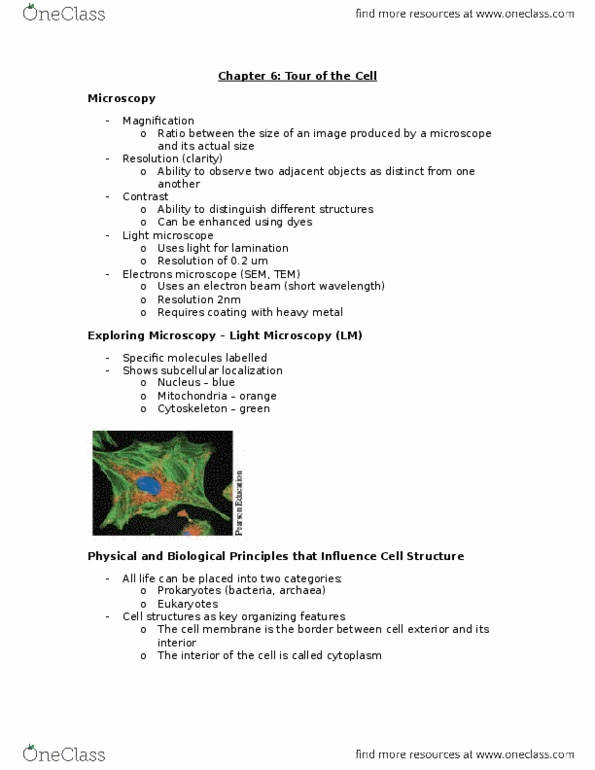
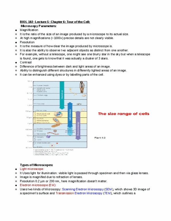
Watch: inner life of a cell (narrated version) Magnification ratio between the size of an image produced by a microscope and its actual size. Resolution ability to observe two adjacent objects as distinct. Contrast ability to distinguish different structures: enhanced using dyes. Light microscope: uses light for illumination, resolution of 0. 2 nanometers. Electron microscope (sem, tem: uses an electron beam with a short wavelength, resolution of 2nm, requires coating with heavy metal (cannot be used on live tissue without damaging it) Dyes light up specific structures of a cell. There is an excitation wavelength and an emission wavelength: the optics is focused on the emission wavelength. Fusion proteins can glow: the cell is essentially making its own dye. Subcellular localization can produce a 3d image. Confocal fluorescence microscopy for sub-cellular 3d models: the laser excites a particular structure that has been stained beforehand (or where a protein has targeted, the laser scans the image and then reproduces a 3d image.