ANAT 101 Lecture Notes - Lecture 16: Bronchiole, Internal Intercostal Muscles, Chronic Obstructive Pulmonary Disease

12 Feb 2015
School
Department
Course
Professor
Document Summary
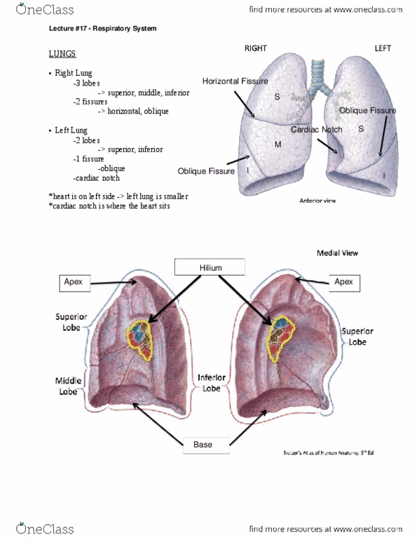
The mediasinum: not a region, central region within the thoracic cavity, contains visceral (organs and important structures outside the lungs, separates the two pleural cavities. Lung structure: costal surface borders inner ribcage, mediastinal borders mediasinum, diaphragmatic surface anterior surface bordering diaphragm. Low for expansion during respiration: fill the lower portion of the ribcage (the pleural cavity) where the lungs end. Left lung: cardiac notch accommodates heart, since it sits a bit left of the middle, lingula little tongue , smaller than right lung. Lobes and fissures: right, 3 lobes (superior, middle, inferior, correlates to the 3 secondary bronchii, horizontal fissure, oblique fissure. Left: 2 lobes (superior and inferior, correlates to the 2 secondary bronchi, oblique fissure. Lung tissue supplied by a given tertiary bronchus. Medial view: hila - area at which important vessels attach to the organ, pulmonary artery and vein, bronchi.