ANAT 101 Lecture Notes - Lecture 12: Thoracic Inlet, Thoracic Vertebrae, Suprasternal Notch

3 Feb 2018
School
Department
Course
Professor
Document Summary
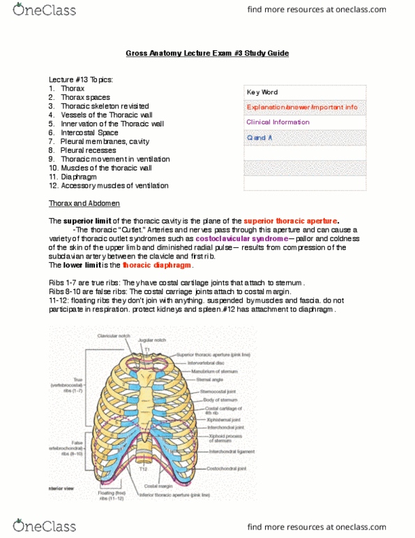
Relationship between thoracic cage and internal organs. Skeletal components of thorax and how they fit together: ribs, sternum, vertebrae. Major muscles associated with thorax make up musculoskeletal wall: breathing mechanics how thorax helps us breathe. Make up whole thorax, people mistakenly call just the rib cage the thoracic cavity. Superior thoracic aperture: first vertebrae, manubrium, first rib. Anterior thoracic aperture: bound by diaphragm from rest of body. Protect vital organs in thorax and in abdominal cavity: diaphragm goes into ribcage, stomach (left) and liver (right) sit under ribcage. Acts as a conduit between head and neck, thorax, abdomen: allows muscles to be connected all the way down. 3 main types of bones: sternum, 12 pairs of ribs, 12 thoracic vertebrae (t1 to t12) Jugular notch right in center, the divot right under our neck, where the clavicle meets with manubrium, the clavicular notch. Move inferior and laterally, meet 1st costal notch, first rib joins sternum.