NURSING 1J02 Lecture Notes - Lecture 7: Abdominal Cavity, Abdominal Wall, Pelvic Examination

8 Mar 2018
School
Department
Course
Professor
Document Summary
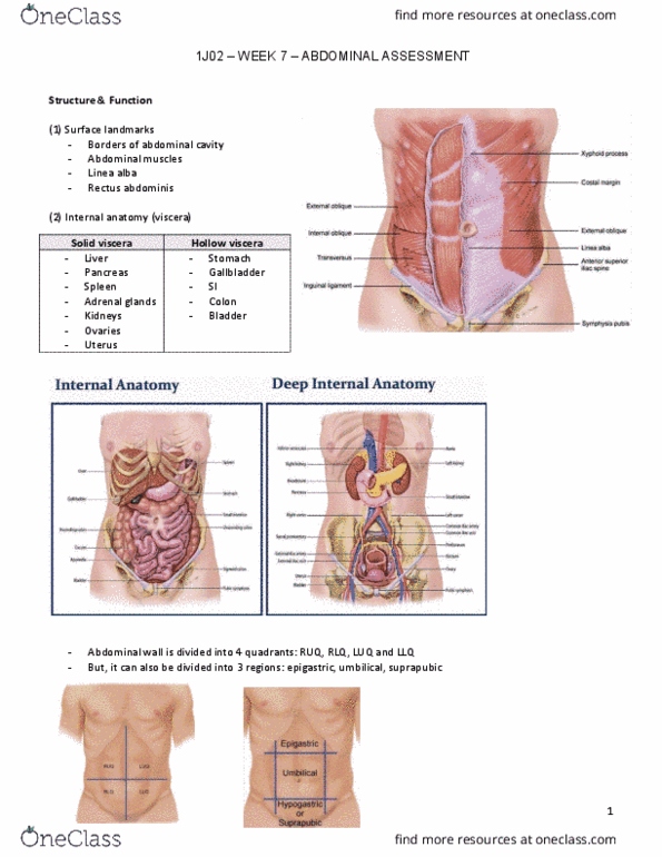
Review structure and function of gi system and abdomen. Large, oval cavity extending from diaphragm to top of pelvis. Bordered in back by vertebral column and paravertebral muscles and at sides and front by lower rib cage and abdominal muscles. 4 layers of large, flat muscles form ventral abdominal wall. Joined at midline by tendinous seam, linea alba. Rectus abdominis forms a strip extending length of midline. Muscles protect and hold organs in place as well as flex vertebral column. Viscera: all organs inside the abdominal cavity. Must be able to visualize where each organ is inside the abdominal cavity. Liver (fills most of the right upper quadrant, and extends over to left midclavicular line) Pancreas - soft, lobulated gland located behind the stomach, in left upper quadrant. Spleen - soft mass of lymphatic tissue on posterolateral wall of abdominal cavity, immediately under the diaphragm. Kidneys - bean shaped, posterior to abdominal contents, left kidney lies at.