EXCI 254 Lecture Notes - Lecture 4: Epidural Space, Jugular Foramen, Epidural Hematoma

4 Feb 2017
School
Department
Course
Professor
Document Summary
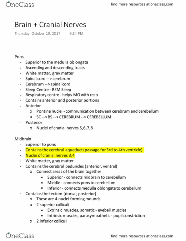
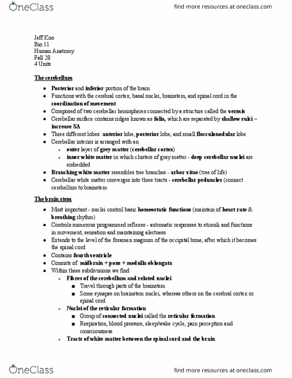
The cerebral peduncles are connecting the spinal cord to the brain. Two parts of rhombencephalon: metencephalon and myelencephalon. Metencephalon has the pons (mostly tracts, white matter. Cerebellum is inferior to the occipital lobe. The cerebellum itself has 2 hemispheres separated by falx cerebelli . Middle line of the cerebellum is the vermis, structure of tracts, white matter. If we dissect cerebellum, sagittal section we see white matter in central part called arbor vitae, these are tracts. The gyri are called the folia and the sulci are are simply the sulci. Folia and sulci creates a finger fimbriae. Cerebellar peduncles, looking from inferiorly at cerebellum, white matter that will bring out the neural pathways. Medulla oblongata is inferior to the pons. What we see at medulla is part of the myelencephalon, 4th ventricle. There ar 2 lateral foramen, foramen of luschka or lateral foramen. Could have csf flow into su(cid:271)ara(cid:272)h(cid:374)oid spa(cid:272)e there.