NEUR30003 Lecture Notes - Lecture 19: Medial Rectus Muscle, Inferior Rectus Muscle, Frontal Eye Fields

18 Jun 2018
School
Department
Course
Professor

Lecture 19
-1. Eye movements are characterised by fixations (centring the foveal part of the retina on the location of
interest) separated by saccades – we don’t acquire any visual information during saccades (retinal
images during saccades would be extremely motion-blurred). Images need to be stability projected onto
the retina, but that stability needs to be fleeting: image drift and continual micro-saccades (minutes of
arc) keep refreshing the activity of photoreceptors.
-2. There are two processes that keep moving images stabilised when the visual target, or our head, is
moving.
- i) The optokinetic response: smooth pursuit of a continually moving scene (e.g. a river or a train passing
in front of us) requires processing by the visual system from which the rate of movement (in angular
terms) is calculated and translated to the same rate of eye rotation – when the extremes of eye
movement in the orbit is reached, a saccade occurs - involuntarily) – this smooth drifting and saccadic
re-centering is called nystagmus (technically, physiological nystagmus, unlike the various forms of
pathological nystagmus seen after nervous system injury).
- ii) The vestibulao-occular response: keeping a central located object foveated while the head is being
rotated requires that sensory signals from the semicircular canals produces the same speed (but
opposite direction) of rotation of the eyeballs.
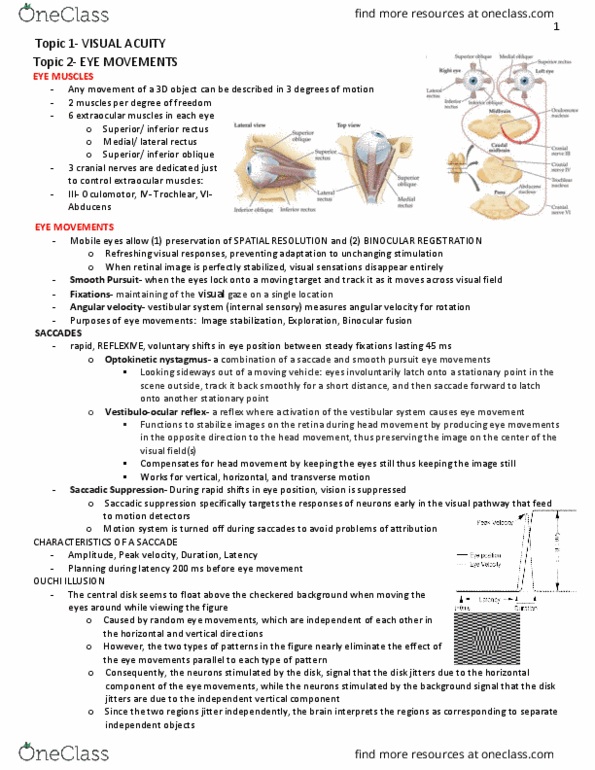
-3. The eye ball’s movement is controlled by 6 muscles; up and down force is applied by the superior and
inferior rectus muscles; side to side by the medial and lateral rectus muscles, and the superior and
inferior oblique pull the eyeball down and in, and up and out respectively. The eye muscles are
innervated by lower motoneurons in three regions of the brainstem and send their axons to the orbit via
their different cranial nerves. In addition to innervating medial, superior and inferior rectus, and inferior
oblique, cranial nerve III also innervate the upper eyelid and carries parasympathetic efferents that
constrict the pupil and accommodates the lens.
-4. Eye muscles work in concert, the three regions of LMNs are connected by the medial longitudinal
fasciculus. Eye movements are ballistic (feed-forward, no feedback mechanism are used to acquire
targets – correcting errors requires new saccade). The cerebellum sets gain of smooth pursuit (and
saccades, presumably).
-5. The firing of LMN causes contraction of the muscle and movement of the eye, but the eyeball will
have a tendency to return to the “neutral” position neural control of eye movement. The burst signals
need to be integrated to maintain new position (ie oppose drift back to neutral) – this happens by action
of cerebellar, pontine circuit neurons. These are divided into a midbrain “vertical gaze centre” and a
pontine “horizontal gaze centre”)
-6. Superior colliculus and frontal eye fields (FEF) represent a map of eye movement vectors: micro-
stimulation produces a movement of particular direction and distance (angle). Experiments have shown
that these neural centres encoding patterns movement that achieve a goal (like foveating on a target
region), not individual movements. Both regions (SC and FEF) exert complimentary (and compensatory
after injury) paths to brainstem gaze control centres.
-7. The single FEF unit fires at different rates - some form of lateral inhibition is evident; the target
(selected for a saccade) is “contrast enhanced” relative to nearby distractors, which are suppressed
more than remote distractors. FEF is involved (along with SC) in smooth pursuit as well as saccades.
FEF gets input from “dorsal stream” of visual information.
-8. The basal ganglia are interconnected with the FEF cortex. Presumably, the basal ganglia are required
for the selection and initiation, and ongoing improvement, of eye movements.
-
- Visual system has low temporal resolution, can’t detect changes that occur rapidly
- When making a saccade → huge blur, can’t keep up with rate of which info is
changing on retina from one point to another
find more resources at oneclass.com
find more resources at oneclass.com

- Visual input is suppressed while saccade is happening
-
- Lateral and medial - side to side
- Superior and inferior - up and down
- Oblique - converge at midline, pull eyeball around middle
- For most of vision is conjugate
- For near vision is disjoint, disconjugate
-
- If fixate on one point → doing a lot of small saccades, Image will drift → then eye will
make small saccade
- Stable retina image → retina loses interest, images becomes grey quickly
find more resources at oneclass.com
find more resources at oneclass.com

-
-
-
find more resources at oneclass.com
find more resources at oneclass.com
Document Summary
The eye muscles are innervated by lower motoneurons in three regions of the brainstem and send their axons to the orbit via their different cranial nerves. Eye movements are ballistic (feed-forward, no feedback mechanism are used to acquire targets correcting errors requires new saccade). The burst signals need to be integrated to maintain new position (ie oppose drift back to neutral) this happens by action of cerebellar, pontine circuit neurons. Experiments have shown that these neural centres encoding patterns movement that achieve a goal (like foveating on a target region), not individual movements. Fef is involved (along with sc) in smooth pursuit as well as saccades. Fef gets input from dorsal stream of visual information: the basal ganglia are interconnected with the fef cortex. Presumably, the basal ganglia are required for the selection and initiation, and ongoing improvement, of eye movements. Visual system has low temporal resolution, can"t detect changes that occur rapidly.