BIOL10002 Lecture Notes - Lecture 14: Pulmonary Valve, Pulmonary Vein, Hemolymph


Lecture 14: Vertebrate heart structure & function
3 weeks after fertilization →beating heart
Main functions of mammalian circulatory system: transport nutrients, oxygen & hormones, and remove metabolic waste
Why do animals need a circulatory system? Circulatory systems evolved along with increased metabolic demands in more complex and larger
animals
● O2, nutrients must be transported around the body to tissues and organs, waste products must be removed
● Communication via hormones, temperature regulation and reproduction
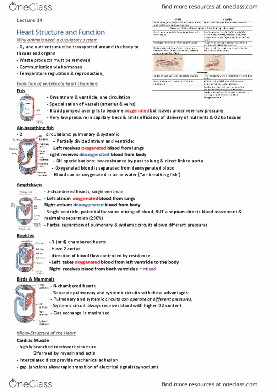
Open circulatory system:
Blood (hemolymph) flows freely within body cavities
making direct contact with all tissues and organs
Heart pumps blood into body cavities through open-
ended vessels→haemolymph flows out and bathes
in tissues
All components of haemolymph leaves the vessels
Fluid drains back to heart; enters when relaxed via
ostia (opening) which acts as valves to ensure one
way fluid
Closed circulatory system:
Heat pumps blood contained in vessels to different regions of the body
Specific components of the fluid filter out of the vessels (in capillaries) to penetrate
tissues; small solutes and water leave and larger molecules & blood cells remain
Transport fluid (blood) is kept separate from fluid that surrounds cells (interstitial fluid)
Fluid returns to the heart via veins, with valves to ensure one way flow of fluid
Advantages:
● Faster transport and more efficient delivery of fluid to tissues
● Ability to control distribution of blood to specific tissues by changing vessel
resistance
● Assists in the delivery of larger molecules (i.e. nutrients and hormones) to
specific tissues
● Enabled the evolution of circulatory systems which keep oxygenated blood
separate from deoxygenated blood
Evolution of vertebrate heart chambers:
Fish: one atrium, one ventricle, one circulation; specialization of vessels
(arteries and veins); blood pumped over gills to become oxygenated but
leaves under very low pressure; very low pressure in capillary beds and
limits efficiency of delivery of nutrients and O2 to tissues
Air-breathing fish: 2 circulations: pulmonary and systemic; partially
divided atrium and ventricle: left receives oxygenated blood from lungs
and right receives deoxygenated blood from body; gill specializations for
low resistance by pass to lung and direct link to aorta; oxygenated
blood is separated from deoxygenated blood; blood can be oxygenated
in air or water
Amphibians: 3-chambered hearts, left atrium for oxygenated blood from lungs; right atrium for deoxygenated blood from body; single ventricle:
potential for some mixing of blood but a septum directs blood movement and maintains separation; partial separation of pulmonary and systemic
circuits allows different pressures
find more resources at oneclass.com
find more resources at oneclass.com

Reptiles: 3 or 4 chambered hearts; have two aortae: left takes
oxygenated blood from left ventricle to the body, right receives blood
from both ventricles (mixed); right aorta (deoxygenated) receives blood
from both ventricles and transports a mix of deoxygenated and
oxygenated blood to the capillaries; reptiles don’t always breathe, blood
by-passes lungs and flows directly to the systemic circuit via the right
aorta - shunt; direction of blood flow is controlled by resistance in the
pulmonary circuit (lower when animal is breathing)
Birds & mammals: 4 chambered hearts; separate pulmonary and
systemic circuits with advantages: pulmonary and systemic circuits can
operate at different pressures, systemic circuit always receives blood
with higher O2 content, gas exchange is maximized
Atrial septal defect (ASD): hole in the septum between atria; congenital,
deoxygenated and oxygenated blood mix and the heart doesn’t work
efficiently; decreased O2 levels in blood, right heart enlargement and
heart failure, pulmonary hypertension (high blood pressure in arteries
supplying the lungs); shortness of breath, fainting, irregular heart
rhythm etc; surgery for treatment
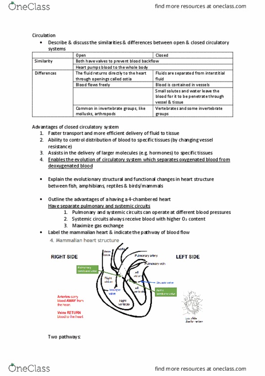
Congenital: present at birth
Apex: bottom, inferior portion of the heart
Vena cava: receives deoxygenated blood
Aorta: sends oxygenated blood to body (systemic circuit)
Pulmonary artery: sends deoxygenated blood to lungs (pulmonary
circuit)
Pulmonary vein: receives blood from lungs (oxygenated); only vein that
carries oxygenated blood
Atrioventricular valves: lie between the atria and ventricles and prevent
backflow into atria when ventricles contract
● Bicuspid/mitral: left
● Tricuspid: right
find more resources at oneclass.com
find more resources at oneclass.com

Atrioventricular groove: runs across top (base) of heart; mainly filled
with fat
Interventricular groove: runs between left and right ventricles,
diagonally up across the heart, mainly filled with fat
Interatrial septum: divides the right and left atrium and contains a thin
area (fossil ovalis); before birth allows blood to flow through a hole
(foramen ovale) to bypass lung
Fossa ovalis: left from the hole (foramen ovale) that existed before birth
to bypass the lungs; created when this hole becomes sealed after birth;
thin wall
Coronary sinus: posterior to fossa ovalis; the main vessel returning
blood from the heart wall
Semilunar valves: pulmonary valve and aortic valves lie between the
ventricles and the major arteries and prevent backflow into ventricles
when the ventricles relax
● Valves prevent backflow
● Blood flows high to low pressure
● Veins bring blood back to heart, arteries take blood away
Septomarginal trabecula: between the interventricular septum and
ventricle wall; has a function in electrical conduction
Chordae tendineae: thin, strings that attach the valve flaps to papillary
muscles on the ventricle walls
Papillary muscle: muscles located in the ventricles of the heart that
attach to the cusps of the AV valves via the chordae tendineae and
contract to prevent inversion or prolapse of these valves
Cardiac muscle:
● Cardiomyocytes: cardiac muscle cells: branched with cross
striations formed by myosin and actin
● Intercalated discs: connect cardiomyocytes, provide
mechanical adhesion & synchronized contraction of cardiac
tissue
● Gap junctions allow rapid & direct transmission of electrical
signal (syncytium) across chambers of the heart; protein-lined
tunnels
● Myocardium: muscular tissue of the heart; the thickness
affects the pressure generated
Heart murmurs: narrowing or leaking of valves (e.g. mitral
regurgitation); congenital, age-related changes, infections, etc; increases
heart work and decreases efficiency, may decrease O2 levels in the
blood; sounds: whooshing, swishing, galloping sounds; asymptomatic,
shortness of breath, pain, fainting etc; valve repair surgery only option if
condition is serious
Diastole & systole refer to what the ventricles are doing: diastole =
relaxation, systole = contraction, therefore when ventricles are relaxed,
atria contract and pump blood into ventricles (diastole)
Diastole: the phase of the heartbeat when the heart muscle relaxes and
allows the chambers to fill with blood
Systole: the last stage of a heartbeat; heart refills with blood; the heart's
two ventricles contract
find more resources at oneclass.com
find more resources at oneclass.com
Document Summary
Main functions of mammalian circulatory system: transport nutrients, oxygen & hormones, and remove metabolic waste. Circulatory systems evolved along with increased metabolic demands in more complex and larger animals. O2, nutrients must be transported around the body to tissues and organs, waste products must be removed. Communication via hormones, temperature regulation and reproduction. Heat pumps blood contained in vessels to different regions of the body making direct contact with all tissues and organs. Heart pumps blood into body cavities through open- ended vessels haemolymph flows out and bathes in tissues. Specific components of the fluid filter out of the vessels (in capillaries) to penetrate tissues; small solutes and water leave and larger molecules & blood cells remain. Transport fluid (blood) is kept separate from fluid that surrounds cells (interstitial fluid) Fluid returns to the heart via veins, with valves to ensure one way flow of fluid.