PHTY208 Lecture Notes - Lecture 13: Pleurisy, Viscosity, Night Sweats


Paediatric pulmonary physiology:
Lung development:!
Embryonic period (0-7 weeks): !
-Primitive lung bud- lung arises as outpouching of gut,main and lobar bronchi form!
-Pulmonary vasculature (arteries and veins) develops and divides with lung in a caudal direction!
Pseudoglandular phase (5-17 weeks):!
-Airway multiplication- bronchial branching and formation completed, > 16 weeks increase in
length and size!
-Formation of muscle fibre, elastic, early cartilage within the bronchi, and mucous glands!
-Diaphragm develops- diphragmatic hernias arise in this timeframe!
Cannicular phase (13-27 weeks):!
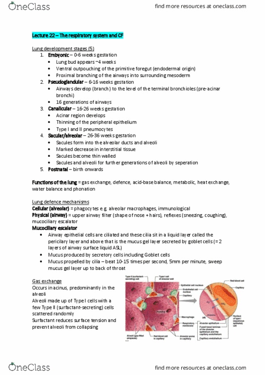
-Development of and vascularisation of respiratory portion of lung- cilia appear on epithelial cells
of trachea and main bronchi, spread towards the periphery, formation of alveolar buds and
sacculi !
-Surfactant production may begin at this phase- also involves cell types which produce the
phospholipid lecithin!
Terminal sac period (24-40 weeks):!
-The phase of cilia, surfactant and alveoli development!
-Terminal sacsa appear as outpouchings of terminal bronchioles!
-Over the next few weeks these multiply forming multiple pouches off the alveolar chamber
(alveolar duct)!
-Epithelial cells differentiate- type 1 cells (95%)- surfactant epithelium thins as vascular
proliferation ^ (creation of the future blood gas barrier), type 2 (5%)- surfactant production!
-At term primitive alveoli are detected!
-Surfactant production- surface active material that reduces surface tension at air liquid
interfaces, present from 18-24 weeks gestation, enables the lungs to remain aerated upon
expiration, preterm babies commonly suffer surfactant insufficiency!
At Birth:!
-Lungs are a heterogenous mix of undifferentiated and differentiated tissue which will develop
and grow- lung, nerves, lymph, blood vessels!
-Alveoli- approx. 20-70 million terminal alveoli, shallow and wider mouth in newborn, change
after few months !
Postantal lung development:!
-Lung development continues after birth, alveoli continue to form until 8-11 years, the number of
generations of bronchi increases from 16 divisions at birth to 23 divisions at 14 years!
Anatomical Differences:!
Airway diameter and length:!
-Narrow airways, smaller diameter, increased resistance to flow!
Heart size:!
-Adult 1/ of rib cage, infant 1/2 of rib cage, less space for lungs!
Chest wall:!
-Soft, compliant chest wall (cartilaginous and therefore retraction), rib cage configuration
(horizontal ribs weak, poorly developed intercostals)!
Diaphragmatic breathing:!
-Horizontal angle of insertion, combined with compliant ribs (less efficient ventilation, distortion
of chest wall shape on inspiration), pattern is piston, not bucket handle, greater susceptibility to
respiratory compromise by things that limit diaphragm movement (feeds, abdominal distention,
head down tips)!
Preferential nasal breathing:!
-Direct airway from nares to lungs, obstruction compromises ventilation, issues with feeding
(nutrative suck-swallow breathe control, non-nutrative suck-breathe control , gastro-
Oesophageal reflux (GOR) and airway damage due to regurgitation)!
Bronchial wall structure:!
-Cartilage less firm , airways prone to collapse easily with neck hyperflexion, hyperextension or
rotation!
Alveoli:!
find more resources at oneclass.com
find more resources at oneclass.com

-Full term infant has no alveoli but 150 million saccule, alveoli do not develop until months old,
full compliment (3-400 million) by 8 years old, decreased surface area for gas exchange!
Physiological differences:!
Lung compliance:!
-Decreased lung compliance, greater inflation pressures are required to maintain lung volumes,
during respiratory distress intercostal, subcostal ands eternal recession may be observed!
Respiratory muscle fatigue:!
-diaphragm primarily type 1 (ST) muscle fibres (adults 55%), 8/12 50%, 3/12 30%, newborn
25%, preterm 10%!
Respiratory rate:!
-Preterm infant 50-70bpm, infant 40bpm, children 18-30bpm, adult 12-15bpm!
-Respiratory compromise - increased respiratory rate, rather than depth of diaphragmatic
excursion!
Sleep state:!
-Neonates sleep up to 20hrs/day, REM sleep (Irregular respiration, loss of postural tone in
muscles, lung volume decreases by 30%, apnoea), adults spend 20% in REM sleep, preterms
spend 80% in REM sleep!
Periodic respiration:!
-Preterm less than 35/40, brief apnoeas of 10-20 seconds, respiratory centre immaturity, worse
with frequent handling!
Increased metabolic rate:!
-neonate has oxygen consumption (newborn 2x an adult), neonate WOB- 40%, adult WOB-
1-2% at rest; 10-20% during exercise, infants will progress to hypoxaemia more rapidly!
Collateral ventilation:!
-Pores of Kohn- intra-alveolar (1-2 years), canals of Lambert- bronchiole- alveolar (6 years),
channels of Martin- inter bronchiolar, late development!
Preferential ventilation:!
-Infants ventilate uppermost lung??, adults ventilate lower (dependent) lung!
Lung volumes:!
-Small VT (closing capacity may impede on tidal volume breathing, results in airway closure in
dependent regions!
Fetal Haemoglobin:!
-Higher Hb concentration in fatal blood > higher affinity for O2, O2 dissociation curve shifts to L!
Muco-ciliary function:!
-Poorly developed mucociliary escalator , increased density of mucous glands, cilial function
impairment !
Immature cough and gag reflexes:!
-Develops at 32-34 weeks, inconsistent until 37 weeks, fatigues easily due to immature
musculature!
Hypothermia:!
-Minimal subcutaneous fat, relatively large surface area mature thermo-regulation mechanism
(affects respiratory drive, affects oxygen consumption)!
Clinical Implications:!
-Increased risk of atelectasis (smaller size of alveoli, lack of collateral ventilation)!
-RUL collapse common (angle of bronchus , airway diameter)!
-High resistance to airflow (smaller diameter airways, distal growth of airways lags behind
proximal)!
-Dynamic compression of trachea (weak cartilaginous support)!
-Increased risk of V/Q mismatch (less alveoli = less surface area for gas exchange , ventilation
preferentially distributed to non-dependent lung regions, perfusion preferentially distributed to
dependent lung regions)!
-Minor insult results in major increase in WOB (^ metabolic rate, angle of ribs, horizontal
insertion of diaphragm, smaller airways, only able to increase rate not depth of respiration)!
Respiratory distress clinical signs:!
-Increased respiratory rate, grunting on expiration, cyanosis, head bobbin, apnoea !
find more resources at oneclass.com
find more resources at oneclass.com

Disorders of Ventilation (part 1):
Pathology:!
-The interruption, cessation, disorder or disease of a body system or organ structure!
Aetiology:!
-The cause, set of causes, or manner of causation of a disease or condition!
Pathogenesis:!
-The sequence of cellular and tissue events that take place from the time of initial contact with
an etiological agent until the ultimate expression of the disease i.e. how the disease process
evolves!
Pathophysiology:!
-The study of the body’s response to dysfunction or disease!
Clinical manifestations:!
-Symptoms- patient reported subjective outcomes !
-Signs- objective outcomes measured by observations, examination, testes, scales!
Airways defence mechanisms:!
Normal airways defence mechanisms!
-Lungs are usually sterile!
-Upper airway is colonised by benign anaerobic bacteria which limit invasion by more
pathogenic bacteria!
3 major risks to the respiratory system:!
1. Inspired air!
2. Pulmonary circulation !
3. Flooding of the alveoli!
Mechanical anatomical defences:!
-Nasal passages- filter and humidify air!
-Upper airway reflexes- epiglottis and bulbar muscles minimise aspiration!
-Mucocillary escalator- mucous glycoprotein produced by goblet cells, trap particles, traps and
clears foreign particles!
-Cough- clear mucous and particles from lower airways!
-Lymphatics- drain fluid from interstitial and assist immune response!
Cellular lung defences!
-Alveolar macrophages reside in alveoli and survey alveolus for foreign material which they
internalise, phagocytose and inactivate. They also release chemical mediators to recruit
neutrophils!
-Neutrophils increase phagocytic activity due to large numbers that are rapidly recruited!
-Natural killer lymphocytes target virally infected and other non-self cells for apoptosis by
releasing membrane permeabilising enzymes !
Molecular lung defences!
-Include cytokines, prostaglandin E2, cell wall hydrolases, iron binding proteins, complement
components, defensives, antiproteinases, surfactant proteins and antioxidants!
-Surfactant- protects alveoli from collapse according to Laplace’s Law P=4T/r!
Infective disorders:!
Upper respiratory tract infection (URTI):!
Definition:!
-Infection of the respiratory system involving anatomical structures including the larynx and
above e.g. the common cold!
Aetiology:$
- Usually viral (=- bacterial)!
Pathogenesis:!
-immune and inflammatory response!
Clinical manifestations:!
-increased mucus production!
-Constitutional symptoms (see acute bronchitis)!
Acute Bronchitis:!
Definition:!
-Reversible bronchial inflammation and mucus production!
find more resources at oneclass.com
find more resources at oneclass.com
Document Summary
Primitive lung bud- lung arises as outpouching of gut,main and lobar bronchi form. Pulmonary vasculature (arteries and veins) develops and divides with lung in a caudal direction. Airway multiplication- bronchial branching and formation completed, > 16 weeks increase in length and size. Formation of muscle bre, elastic, early cartilage within the bronchi, and mucous glands. Diaphragm develops- diphragmatic hernias arise in this timeframe. Development of and vascularisation of respiratory portion of lung- cilia appear on epithelial cells of trachea and main bronchi, spread towards the periphery, formation of alveolar buds and sacculi. Surfactant production may begin at this phase- also involves cell types which produce the phospholipid lecithin. The phase of cilia, surfactant and alveoli development. Terminal sacsa appear as outpouchings of terminal bronchioles. Over the next few weeks these multiply forming multiple pouches o the alveolar chamber (alveolar duct)