NSG 2317 Chapter Notes - Chapter 21: Intercostal Space, Ileocecal Valve, Stethoscope

15 Dec 2016
School
Department
Course
Professor
Document Summary
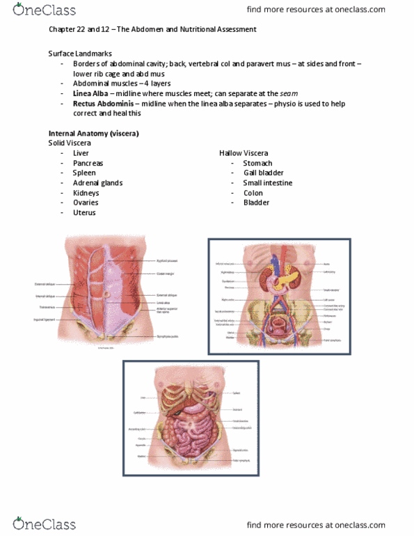
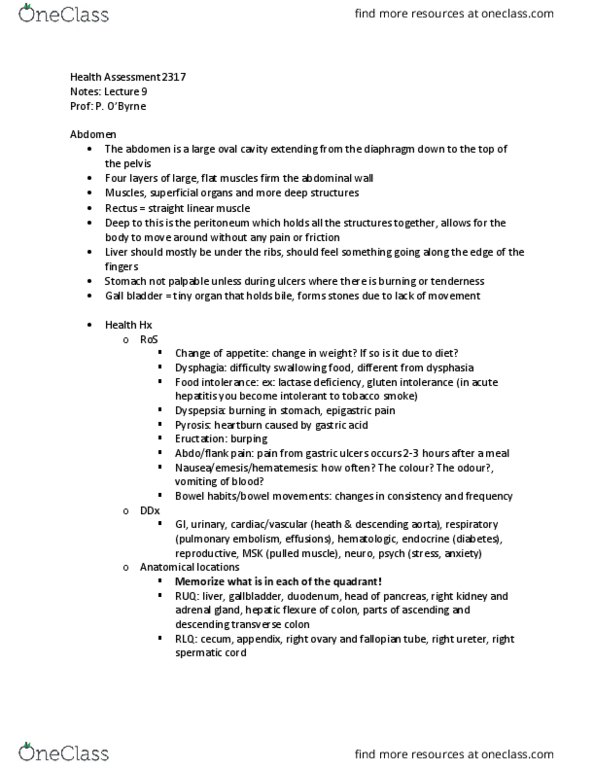
Chapter 21 - the abdomen: structure and function, subjective data health history questions, objective data the physical exam, abnormal findings. Abdomen: large oval cavity extending from diaphragm down to top of the pelvis. Abdominal cavity bordered by vertebral column & paravertebral muscles at side & front by lower rib cage & abdominal muscles. 4 large, flat layers of muscle form ventral abdominal wall, joined by linea alba & rectus abdominis (often palpable) Solid viscera: maintain a characteristic shape: liver: most of ruq; normally palpable, pancreas, spleen, adrenal glands, kidneys, ovaries: normally palpable only on bimanual assessment during pelvic exam, uterus. Spleen: soft mass of lymphatic tissue on the posterolateral wall of abdominal wall, under diaphragm. Aorta: upper middle part of abdomen, bifurcates into right & left common palpable. Subjective data health history questions: appetite, dysphagia, food intolerance, abdominal pain, nausea/vomiting, bowel habits, abdominal history, medications, nutritional assessment.