BIOL 171 Study Guide - Midterm Guide: Longus Colli Muscle, Internal Jugular Vein, Jugular Vein

Document Summary
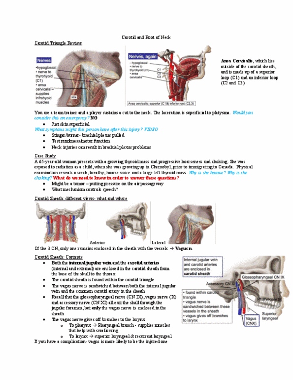
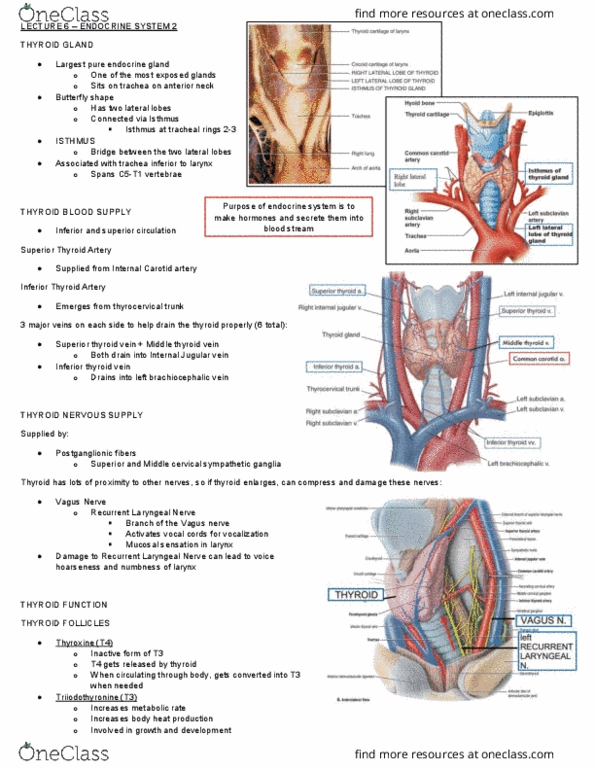
The two lobes (right and left) of the thyroid are connected by. Isthmus: a thin, hypoechoic band immediately anterior to each thyroid lobe, the sternocleidomastoid muscle lies anterior and lateral to the thyroid. What structures are part of the major neurovascular bundle: carotid sheath (carotid artery, internal jugular vein and vagus nerve) Longus colli muscle: vagus nerve, carotid artery. How does the normal thyroid look sonographically: normal thyroid tissue demonstrates a fine homogenous. How do the muscles around the thyroid look sonographically? echo texture: the surrounding muscles appear hypoechoic to the thyroid gland. Length: 4-6 cm: anteroposterior: 1. 5 to 2. 0 cm in width. What is the landmark for the posterolateral border of the thyroid in long axis: common carotid artery. ______ of the population has a pyramidal lobe: pyramid lobe, 15-30% What are the thyroid hormones: t3 = triiodothyronine plays a key role in metabolic. Calcitonin is secreted by ______ of the thyroid gland and.